 Movie
Movie Controller

[English] 日本語
 Yorodumi
- PDB-8b72: Crystal structure of 3-hydroxydecanoyl-acyl carrier protein dehyd... -
+ Open data

- Basic information
| Entry | Database: PDB / ID: 8b72 | ||||||
|---|---|---|---|---|---|---|---|
| Title | Crystal structure of 3-hydroxydecanoyl-acyl carrier protein dehydratase (FabA) from Pseudomonas aeruginosa in complex with Z30857828 | ||||||
 Components Components | 3-hydroxydecanoyl-[acyl-carrier-protein] dehydratase | ||||||
 Keywords Keywords | LYASE / FABA / PA1610 | ||||||
| Function / homology |  Function and homology information Function and homology informationtrans-2-decenoyl-[acyl-carrier protein] isomerase / trans-2-decenoyl-acyl-carrier-protein isomerase activity / unsaturated fatty acid biosynthetic process / 3-hydroxyacyl-[acyl-carrier-protein] dehydratase / (3R)-hydroxyacyl-[acyl-carrier-protein] dehydratase activity / fatty acid biosynthetic process / cytosol Similarity search - Function | ||||||
| Biological species |   Pseudomonas aeruginosa (bacteria) Pseudomonas aeruginosa (bacteria) | ||||||
| Method |  X-RAY DIFFRACTION / SYNCHROTRON / MOLECULAR REPLACEMENT / Resolution: 1.87 Å X-RAY DIFFRACTION / SYNCHROTRON / MOLECULAR REPLACEMENT / Resolution: 1.87 Å | ||||||
 Authors Authors | Robinson, D.A. / Moynie, L. / Naismith, J.H. / Gray, D.W. | ||||||
| Funding support |  United Kingdom, 1items United Kingdom, 1items
| ||||||
 Citation Citation | Journal: To Be Published Title: Crystal structure of 3-hydroxydecanoyl-acyl carrier protein dehydratase (FabA) from Pseudomonas aeruginosa in complex with Z30857828 Authors: Robinson, D.A. / Gray, D.W.G. | ||||||
| History |
|
- Structure visualization
| Structure viewer | Molecule: MolmilJmol/JSmol |
|---|
- Downloads & links
-Download
| PDBx/mmCIF format | 8b72.cif.gz | 186.7 KB | Display | PDBx/mmCIF format |
|---|---|---|---|---|
| PDB format | pdb8b72.ent.gz | 148.6 KB | Display | PDB format |
| PDBx/mmJSON format | 8b72.json.gz | Tree view | PDBx/mmJSON format | |
| Others |  Other downloads Other downloads |
-Validation report
| Arichive directory | https://data.pdbj.org/pub/pdb/validation_reports/b7/8b72ftp://data.pdbj.org/pub/pdb/validation_reports/b7/8b72 | HTTPS FTP |
|---|
-Related structure data
| Related structure data | 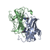 4cl6S S: Starting model for refinement |
|---|---|
| Similar structure data |
-Links
 PDBj
PDBj
- Assembly
| Deposited unit | 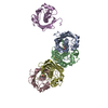
| ||||||||
|---|---|---|---|---|---|---|---|---|---|
| 1 | 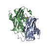
| ||||||||
| 2 | 
| ||||||||
| 3 | 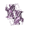
| ||||||||
| Unit cell |
|
-Components
| #1: Protein | Mass: 18768.533 Da / Num. of mol.: 5 Source method: isolated from a genetically manipulated source Source: (gene. exp.) Pseudomonas aeruginosa (bacteria)Strain: ATCC 15692 / DSM 22644 / CIP 104116 / JCM 14847 / LMG 12228 / 1C / PRS 101 / PAO1 Gene: fabA, PA1610 / Production host: References: UniProt: O33877, 3-hydroxyacyl-[acyl-carrier-protein] dehydratase, trans-2-decenoyl-[acyl-carrier protein] isomerase #2: Chemical | ChemComp-M25 / |   Mass: 242.295 Da / Num. of mol.: 1 / Source method: obtained synthetically / Formula: C10H14N2O3S / Feature type: SUBJECT OF INVESTIGATION Mass: 242.295 Da / Num. of mol.: 1 / Source method: obtained synthetically / Formula: C10H14N2O3S / Feature type: SUBJECT OF INVESTIGATION#3: Chemical |   Mass: 78.133 Da / Num. of mol.: 2 / Source method: obtained synthetically / Formula: C2H6OS / Comment: DMSO, precipitant*YM Mass: 78.133 Da / Num. of mol.: 2 / Source method: obtained synthetically / Formula: C2H6OS / Comment: DMSO, precipitant*YM#4: Water | ChemComp-HOH / |  Mass: 18.015 Da / Num. of mol.: 609 / Source method: isolated from a natural source / Formula: H2O Mass: 18.015 Da / Num. of mol.: 609 / Source method: isolated from a natural source / Formula: H2OHas ligand of interest | Y | |
|---|
-Experimental details
-Experiment
| Experiment | Method: X-RAY DIFFRACTION / Number of used crystals: 1 |
|---|
- Sample preparation
| Crystal | Density Matthews: 3.08 Å3/Da / Density % sol: 60.1 % |
|---|---|
| Crystal grow | Temperature: 293 K / Method: vapor diffusion / Details: PEG 4000, Ammonium sulfate, sodium citrate |
-Data collection
| Diffraction | Mean temperature: 100 K / Serial crystal experiment: N |
|---|---|
| Diffraction source | Source: SYNCHROTRON / Site: Diamond / Beamline: I04-1 / Wavelength: 0.91587 Å |
| Detector | Type: DECTRIS PILATUS 6M / Detector: PIXEL / Date: Jan 11, 2018 |
| Radiation | Protocol: SINGLE WAVELENGTH / Monochromatic (M) / Laue (L): M / Scattering type: x-ray |
| Radiation wavelength | Wavelength: 0.91587 Å / Relative weight: 1 |
| Reflection | Resolution: 1.87→71.55 Å / Num. obs: 92452 / % possible obs: 99.7 % / Redundancy: 3.5 % / CC1/2: 0.99 / Rmerge(I) obs: 0.046 / Net I/σ(I): 10.8 |
| Reflection shell | Resolution: 1.87→1.97 Å / Rmerge(I) obs: 1.115 / Mean I/σ(I) obs: 0.9 / Num. unique obs: 13512 / CC1/2: 0.46 |
- Processing
| Software |
| ||||||||||||||||||||||||||||||||||||||||||||||||||||||||||||
|---|---|---|---|---|---|---|---|---|---|---|---|---|---|---|---|---|---|---|---|---|---|---|---|---|---|---|---|---|---|---|---|---|---|---|---|---|---|---|---|---|---|---|---|---|---|---|---|---|---|---|---|---|---|---|---|---|---|---|---|---|---|
| Refinement | Method to determine structure: MOLECULAR REPLACEMENT Starting model: 4CL6 Resolution: 1.87→71.55 Å / Cor.coef. Fo:Fc: 0.969 / Cor.coef. Fo:Fc free: 0.955 / SU B: 4.523 / SU ML: 0.122 / Cross valid method: THROUGHOUT / σ(F): 0 / ESU R: 0.124 / ESU R Free: 0.123 / Stereochemistry target values: MAXIMUM LIKELIHOOD Details: HYDROGENS HAVE BEEN ADDED IN THE RIDING POSITIONS U VALUES : REFINED INDIVIDUALLY
| ||||||||||||||||||||||||||||||||||||||||||||||||||||||||||||
| Solvent computation | Ion probe radii: 0.8 Å / Shrinkage radii: 0.8 Å / VDW probe radii: 1.2 Å / Solvent model: MASK | ||||||||||||||||||||||||||||||||||||||||||||||||||||||||||||
| Displacement parameters | Biso max: 235.91 Å2 / Biso mean: 45.488 Å2 / Biso min: 27.55 Å2
| ||||||||||||||||||||||||||||||||||||||||||||||||||||||||||||
| Refinement step | Cycle: final / Resolution: 1.87→71.55 Å
| ||||||||||||||||||||||||||||||||||||||||||||||||||||||||||||
| Refine LS restraints |
| ||||||||||||||||||||||||||||||||||||||||||||||||||||||||||||
| LS refinement shell | Resolution: 1.873→1.922 Å / Rfactor Rfree error: 0 / Total num. of bins used: 20
|
