 Movie
Movie Controller


+ Open data

- Basic information
| Entry | Database: PDB / ID: 8b65 | |||||||||
|---|---|---|---|---|---|---|---|---|---|---|
| Title | Structure of rsCherry crystallized in anaerobic conditions | |||||||||
 Components Components | rsCherry | |||||||||
 Keywords Keywords | FLUORESCENT PROTEIN / rsCherry / red fluorescent protein / chemical modifications | |||||||||
| Function / homology | Green fluorescent protein, GFP / Green fluorescent protein-related / Green fluorescent protein / Green fluorescent protein / bioluminescence / generation of precursor metabolites and energy / DI(HYDROXYETHYL)ETHER / Red fluorescent protein drFP583 Function and homology information Function and homology information | |||||||||
| Biological species |  Discosoma sp. (sea anemone) Discosoma sp. (sea anemone) | |||||||||
| Method |  X-RAY DIFFRACTION / SYNCHROTRON / MOLECULAR REPLACEMENT / molecular replacement / Resolution: 1.55 Å X-RAY DIFFRACTION / SYNCHROTRON / MOLECULAR REPLACEMENT / molecular replacement / Resolution: 1.55 Å | |||||||||
 Authors Authors | Bui, T.Y.H. / Van Meervelt, L. | |||||||||
| Funding support | Viet Nam,  Belgium, 2items Belgium, 2items
| |||||||||
 Citation Citation | Journal: Int.J.Biol.Macromol. / Year: 2023 Title: Oxygen-induced chromophore degradation in the photoswitchable red fluorescent protein rsCherry. Authors: Bui, T.Y.H. / De Zitter, E. / Moeyaert, B. / Pecqueur, L. / Srinivasu, B.Y. / Economou, A. / Fontecave, M. / Van Meervelt, L. / Dedecker, P. / Pedre, B. #1: Journal: Biorxiv / Year: 2023Title: Oxygen-induced chromophore degradation in the photoswitchable red fluorescent protein rsCherry Authors: Bui, T.Y.H. / De Zitter, E. / Moeyaert, B. / Pecqueur, L. / Srinivasu, B.Y. / Economou, A. / Fontecave, M. / Van Meervelt, L. / Dedecker, P. / Pedre, B. | |||||||||
| History |
|
- Structure visualization
| Structure viewer | Molecule: MolmilJmol/JSmol |
|---|
- Downloads & links
-Download
| PDBx/mmCIF format | 8b65.cif.gz | 74.7 KB | Display | PDBx/mmCIF format |
|---|---|---|---|---|
| PDB format | pdb8b65.ent.gz | 51.3 KB | Display | PDB format |
| PDBx/mmJSON format | 8b65.json.gz | Tree view | PDBx/mmJSON format | |
| Others |  Other downloads Other downloads |
-Validation report
| Arichive directory | https://data.pdbj.org/pub/pdb/validation_reports/b6/8b65ftp://data.pdbj.org/pub/pdb/validation_reports/b6/8b65 | HTTPS FTP |
|---|
-Related structure data
| Related structure data |  8b7gC  2h5qS S: Starting model for refinement C: citing same article ( |
|---|---|
| Similar structure data |
-Links
 PDBj
PDBj

- Assembly
| Deposited unit | 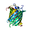
| ||||||||
|---|---|---|---|---|---|---|---|---|---|
| 1 |
| ||||||||
| Unit cell |
|
-Components
-Protein , 1 types, 1 molecules A
| #1: Protein | Mass: 30709.418 Da / Num. of mol.: 1 Source method: isolated from a genetically manipulated source Details: Chromophore structures : QYX, Q2K / Source: (gene. exp.) Discosoma sp. (sea anemone) / Production host:  |
|---|
-Non-polymers , 5 types, 274 molecules 


| #2: Chemical | ChemComp-PEG /  Mass: 106.120 Da / Num. of mol.: 1 / Source method: obtained synthetically / Formula: C4H10O3 Mass: 106.120 Da / Num. of mol.: 1 / Source method: obtained synthetically / Formula: C4H10O3 | ||||
|---|---|---|---|---|---|
| #3: Chemical | ChemComp-MES /  Mass: 195.237 Da / Num. of mol.: 1 / Source method: obtained synthetically / Formula: C6H13NO4S / Comment: pH buffer*YM Mass: 195.237 Da / Num. of mol.: 1 / Source method: obtained synthetically / Formula: C6H13NO4S / Comment: pH buffer*YM | ||||
| #4: Chemical |  Mass: 62.068 Da / Num. of mol.: 2 / Source method: obtained synthetically / Formula: C2H6O2 Mass: 62.068 Da / Num. of mol.: 2 / Source method: obtained synthetically / Formula: C2H6O2#5: Chemical | ChemComp-GOL / |  Mass: 92.094 Da / Num. of mol.: 1 / Source method: obtained synthetically / Formula: C3H8O3 Mass: 92.094 Da / Num. of mol.: 1 / Source method: obtained synthetically / Formula: C3H8O3#6: Water | ChemComp-HOH / | Mass: 18.015 Da / Num. of mol.: 269 / Source method: isolated from a natural source / Formula: H2O |
-Details
| Has ligand of interest | Y |
|---|---|
| Has protein modification | Y |
-Experimental details
-Experiment
| Experiment | Method: X-RAY DIFFRACTION / Number of used crystals: 1 |
|---|
- Sample preparation
| Crystal | Density Matthews: 2.56 Å3/Da / Density % sol: 51.93 % |
|---|---|
| Crystal grow | Temperature: 294.15 K / Method: microbatch / pH: 6 / Details: 18% PEG 8000 and 0.1M MES pH 6 |
-Data collection
| Diffraction | Mean temperature: 100 K / Serial crystal experiment: N | ||||||||||||||||||||||||||||||
|---|---|---|---|---|---|---|---|---|---|---|---|---|---|---|---|---|---|---|---|---|---|---|---|---|---|---|---|---|---|---|---|
| Diffraction source | Source: SYNCHROTRON / Site: SOLEIL  / Beamline: PROXIMA 2 / Wavelength: 0.98 Å / Beamline: PROXIMA 2 / Wavelength: 0.98 Å | ||||||||||||||||||||||||||||||
| Detector | Type: DECTRIS EIGER X 9M / Detector: PIXEL / Date: Jul 12, 2019 | ||||||||||||||||||||||||||||||
| Radiation | Protocol: SINGLE WAVELENGTH / Monochromatic (M) / Laue (L): M / Scattering type: x-ray | ||||||||||||||||||||||||||||||
| Radiation wavelength | Wavelength: 0.98 Å / Relative weight: 1 | ||||||||||||||||||||||||||||||
| Reflection | Resolution: 1.55→46.2 Å / Num. obs: 39472 / % possible obs: 99.6 % / Redundancy: 6.8 % / Biso Wilson estimate: 18.08 Å2 / CC1/2: 0.999 / Rmerge(I) obs: 0.06 / Rpim(I) all: 0.025 / Rrim(I) all: 0.065 / Net I/σ(I): 16.8 / Num. measured all: 267195 / Scaling rejects: 9 | ||||||||||||||||||||||||||||||
| Reflection shell | Diffraction-ID: 1
|
-Phasing
| Phasing | Method: molecular replacement | |||||||||
|---|---|---|---|---|---|---|---|---|---|---|
| Phasing MR |
|
- Processing
| Software |
| |||||||||||||||||||||||||||||||||||||||||||||||||||||||||||||||||||||||||||||||||||||||||||||||||||||||||
|---|---|---|---|---|---|---|---|---|---|---|---|---|---|---|---|---|---|---|---|---|---|---|---|---|---|---|---|---|---|---|---|---|---|---|---|---|---|---|---|---|---|---|---|---|---|---|---|---|---|---|---|---|---|---|---|---|---|---|---|---|---|---|---|---|---|---|---|---|---|---|---|---|---|---|---|---|---|---|---|---|---|---|---|---|---|---|---|---|---|---|---|---|---|---|---|---|---|---|---|---|---|---|---|---|---|---|
| Refinement | Method to determine structure: MOLECULAR REPLACEMENT Starting model: 2H5Q Resolution: 1.55→46.2 Å / SU ML: 0.14 / Cross valid method: THROUGHOUT / σ(F): 1.34 / Phase error: 16.87 / Stereochemistry target values: ML
| |||||||||||||||||||||||||||||||||||||||||||||||||||||||||||||||||||||||||||||||||||||||||||||||||||||||||
| Solvent computation | Shrinkage radii: 0.9 Å / VDW probe radii: 1.11 Å / Solvent model: FLAT BULK SOLVENT MODEL | |||||||||||||||||||||||||||||||||||||||||||||||||||||||||||||||||||||||||||||||||||||||||||||||||||||||||
| Displacement parameters | Biso max: 71.82 Å2 / Biso mean: 22.4133 Å2 / Biso min: 12.06 Å2 | |||||||||||||||||||||||||||||||||||||||||||||||||||||||||||||||||||||||||||||||||||||||||||||||||||||||||
| Refinement step | Cycle: final / Resolution: 1.55→46.2 Å
| |||||||||||||||||||||||||||||||||||||||||||||||||||||||||||||||||||||||||||||||||||||||||||||||||||||||||
| LS refinement shell | Refine-ID: X-RAY DIFFRACTION / Rfactor Rfree error: 0 / Total num. of bins used: 14
|
