 Movie
Movie Controller

[English] 日本語
 Yorodumi
- PDB-8b5t: Crystal structure of Quinonoid dihydropteridine reductase from Le... -
+ Open data

- Basic information
| Entry | Database: PDB / ID: 8b5t | ||||||
|---|---|---|---|---|---|---|---|
| Title | Crystal structure of Quinonoid dihydropteridine reductase from Leishmania major | ||||||
 Components Components | Quinonoid dihydropteridine reductase | ||||||
 Keywords Keywords | OXIDOREDUCTASE / QDPR / LEISHMANIA | ||||||
| Function / homology |  Function and homology information Function and homology information6,7-dihydropteridine reductase / 6,7-dihydropteridine reductase activity / NADH binding / tetrahydrobiopterin biosynthetic process / L-phenylalanine catabolic process / NADPH binding / cytoplasm Similarity search - Function | ||||||
| Biological species |  Leishmania major (eukaryote) Leishmania major (eukaryote) | ||||||
| Method |  X-RAY DIFFRACTION / SYNCHROTRON / MOLECULAR REPLACEMENT / molecular replacement / Resolution: 2.1 Å X-RAY DIFFRACTION / SYNCHROTRON / MOLECULAR REPLACEMENT / molecular replacement / Resolution: 2.1 Å | ||||||
 Authors Authors | Robinson, D.A. / Fairlamb, A.H. | ||||||
| Funding support | 1items
| ||||||
 Citation Citation | Journal: To Be Published Title: Crystal structure of Quinonoid dihydropteridine reductase from Leishmania major Authors: Robinson, D.A. / Fairlamb, A.H. | ||||||
| History |
|
- Structure visualization
| Structure viewer | Molecule: MolmilJmol/JSmol |
|---|
- Downloads & links
-Download
| PDBx/mmCIF format | 8b5t.cif.gz | 139.6 KB | Display | PDBx/mmCIF format |
|---|---|---|---|---|
| PDB format | pdb8b5t.ent.gz | 108 KB | Display | PDB format |
| PDBx/mmJSON format | 8b5t.json.gz | Tree view | PDBx/mmJSON format | |
| Others |  Other downloads Other downloads |
-Validation report
| Arichive directory | https://data.pdbj.org/pub/pdb/validation_reports/b5/8b5tftp://data.pdbj.org/pub/pdb/validation_reports/b5/8b5t | HTTPS FTP |
|---|
-Related structure data
| Related structure data |  1dirS S: Starting model for refinement |
|---|---|
| Similar structure data |
-Links
 PDBj
PDBj


- Assembly
| Deposited unit | 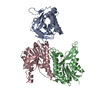
| ||||||||
|---|---|---|---|---|---|---|---|---|---|
| 1 | 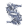
| ||||||||
| 2 | 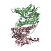
| ||||||||
| Unit cell |
| ||||||||
| Components on special symmetry positions |
|
-Components
| #1: Protein | Mass: 23707.775 Da / Num. of mol.: 3 Source method: isolated from a genetically manipulated source Source: (gene. exp.) Leishmania major (eukaryote)Gene: QDPR-7, QDPR, QDPR-2, QDPR-3, QDPR-4, QDPR-5, QDPR-6, LMJF_34_4360, LMJF_34_4390, LMJF_34_4420, LMJF_34_4450, LMJF_34_4480, LMJF_34_4510 Production host:  References: UniProt: Q4Q290, 6,7-dihydropteridine reductase, EC: 1.6.99.7 #2: Water | ChemComp-HOH / |  Mass: 18.015 Da / Num. of mol.: 362 / Source method: isolated from a natural source / Formula: H2O Mass: 18.015 Da / Num. of mol.: 362 / Source method: isolated from a natural source / Formula: H2O |
|---|
-Experimental details
-Experiment
| Experiment | Method: X-RAY DIFFRACTION / Number of used crystals: 1 |
|---|
- Sample preparation
| Crystal | Density Matthews: 2.44 Å3/Da / Density % sol: 49.58 % |
|---|---|
| Crystal grow | Temperature: 293 K / Method: vapor diffusion / pH: 5.5 / Details: 15% PEG4000, 0.2M MgCl2, 0.1M Na ACETATE pH 5.5 |
-Data collection
| Diffraction | Mean temperature: 100 K / Serial crystal experiment: N |
|---|---|
| Diffraction source | Source: SYNCHROTRON / Site: Diamond  / Beamline: I03 / Wavelength: 0.92045 Å / Beamline: I03 / Wavelength: 0.92045 Å |
| Detector | Type: DECTRIS PILATUS 2M / Detector: PIXEL / Date: Dec 8, 2013 |
| Radiation | Protocol: SINGLE WAVELENGTH / Monochromatic (M) / Laue (L): M / Scattering type: x-ray |
| Radiation wavelength | Wavelength: 0.92045 Å / Relative weight: 1 |
| Reflection | Resolution: 2.1→39.5 Å / Num. obs: 39899 / % possible obs: 99.2 % / Redundancy: 8.4 % / CC1/2: 0.99 / Rmerge(I) obs: 0.072 / Net I/σ(I): 19 |
| Reflection shell | Resolution: 2.1→2.16 Å / Redundancy: 8.8 % / Rmerge(I) obs: 0.781 / Mean I/σ(I) obs: 2.9 / Num. unique obs: 3192 / CC1/2: 0.8 / % possible all: 98.1 |
-Phasing
| Phasing | Method: molecular replacement |
|---|
- Processing
| Software |
| ||||||||||||||||||||||||||||||||||||||||||||||||||||||||||||
|---|---|---|---|---|---|---|---|---|---|---|---|---|---|---|---|---|---|---|---|---|---|---|---|---|---|---|---|---|---|---|---|---|---|---|---|---|---|---|---|---|---|---|---|---|---|---|---|---|---|---|---|---|---|---|---|---|---|---|---|---|---|
| Refinement | Method to determine structure: MOLECULAR REPLACEMENT Starting model: 1DIR Resolution: 2.1→39.5 Å / Cor.coef. Fo:Fc: 0.96 / Cor.coef. Fo:Fc free: 0.931 / SU B: 5.131 / SU ML: 0.133 / Cross valid method: THROUGHOUT / σ(F): 0 / ESU R: 0.216 / ESU R Free: 0.186 / Stereochemistry target values: MAXIMUM LIKELIHOOD Details: HYDROGENS HAVE BEEN ADDED IN THE RIDING POSITIONS U VALUES : REFINED INDIVIDUALLY
| ||||||||||||||||||||||||||||||||||||||||||||||||||||||||||||
| Solvent computation | Ion probe radii: 0.8 Å / Shrinkage radii: 0.8 Å / VDW probe radii: 1.2 Å / Solvent model: MASK | ||||||||||||||||||||||||||||||||||||||||||||||||||||||||||||
| Displacement parameters | Biso max: 155.29 Å2 / Biso mean: 36.974 Å2 / Biso min: 14.11 Å2
| ||||||||||||||||||||||||||||||||||||||||||||||||||||||||||||
| Refinement step | Cycle: final / Resolution: 2.1→39.5 Å
| ||||||||||||||||||||||||||||||||||||||||||||||||||||||||||||
| Refine LS restraints |
| ||||||||||||||||||||||||||||||||||||||||||||||||||||||||||||
| LS refinement shell | Resolution: 2.1→2.155 Å / Rfactor Rfree error: 0 / Total num. of bins used: 20
|
