 Movie
Movie Controller

[English] 日本語
 Yorodumi
- PDB-8b4x: X-ray structure of furin (PCSK3) in complex with Guanidinomethyl-... -
+ Open data

- Basic information
| Entry | Database: PDB / ID: 8b4x | ||||||
|---|---|---|---|---|---|---|---|
| Title | X-ray structure of furin (PCSK3) in complex with Guanidinomethyl-Phac-R-Tle-K-6-(aminomethyl)-3-amino-isoindol | ||||||
 Components Components |
| ||||||
 Keywords Keywords | HYDROLASE / furin / proprotein convertase subtilisin/kexin type 3 / PCSK3 / SARS-CoV-2 / inhibitor / protease / complex | ||||||
| Function / homology |  Function and homology information Function and homology informationfurin / nerve growth factor production / dibasic protein processing / plasma lipoprotein particle remodeling / NGF processing / regulation of cholesterol transport / negative regulation of transforming growth factor beta1 production / Assembly of active LPL and LIPC lipase complexes / : / negative regulation of low-density lipoprotein particle receptor catabolic process ...furin / nerve growth factor production / dibasic protein processing / plasma lipoprotein particle remodeling / NGF processing / regulation of cholesterol transport / negative regulation of transforming growth factor beta1 production / Assembly of active LPL and LIPC lipase complexes / : / negative regulation of low-density lipoprotein particle receptor catabolic process / peptide biosynthetic process / Pre-NOTCH Processing in Golgi / nerve growth factor binding / Synthesis and processing of ENV and VPU / cytokine precursor processing / secretion by cell / Formation of the cornified envelope / Signaling by PDGF / trans-Golgi network transport vesicle / heparan sulfate binding / Signaling by NODAL / Elastic fibre formation / blastocyst formation / peptide hormone processing / positive regulation of membrane protein ectodomain proteolysis / CD163 mediating an anti-inflammatory response / zymogen activation / Regulation of CDH1 posttranslational processing and trafficking to plasma membrane / endopeptidase inhibitor activity / Activation of Matrix Metalloproteinases / Collagen degradation / collagen catabolic process / TGF-beta receptor signaling activates SMADs / Maturation of hRSV A proteins / regulation of protein catabolic process / Respiratory syncytial virus (RSV) attachment and entry / extracellular matrix disassembly / Uptake and function of anthrax toxins / regulation of signal transduction / Removal of aminoterminal propeptides from gamma-carboxylated proteins / endopeptidase activator activity / viral life cycle / extracellular matrix organization / peptide binding / transforming growth factor beta receptor signaling pathway / serine-type peptidase activity / cholesterol homeostasis / serine-type endopeptidase inhibitor activity / trans-Golgi network / negative regulation of inflammatory response to antigenic stimulus / protein maturation / SMAD2/SMAD3:SMAD4 heterotrimer regulates transcription / protein processing / Golgi lumen / peptidase activity / heparin binding / protease binding / endopeptidase activity / Induction of Cell-Cell Fusion / amyloid fibril formation / Potential therapeutics for SARS / positive regulation of viral entry into host cell / Attachment and Entry / endosome membrane / viral protein processing / membrane raft / Amyloid fiber formation / serine-type endopeptidase activity / Golgi membrane / cell surface / endoplasmic reticulum / extracellular exosome / extracellular region / membrane / metal ion binding / plasma membrane Similarity search - Function | ||||||
| Biological species |  Homo sapiens (human) Homo sapiens (human)synthetic construct (others) | ||||||
| Method |  X-RAY DIFFRACTION / SYNCHROTRON / MOLECULAR REPLACEMENT / Resolution: 1.6 Å X-RAY DIFFRACTION / SYNCHROTRON / MOLECULAR REPLACEMENT / Resolution: 1.6 Å | ||||||
 Authors Authors | Dahms, S.O. / Brandstetter, H. | ||||||
| Funding support |  Austria, 1items Austria, 1items
| ||||||
 Citation Citation | Journal: Chemmedchem / Year: 2024 Title: Fragment-Based Design, Synthesis, and Characterization of Aminoisoindole-Derived Furin Inhibitors. Authors: Lange, R.W. / Bloch, K. / Heindl, M.R. / Wollenhaupt, J. / Weiss, M.S. / Brandstetter, H. / Klebe, G. / Falcone, F.H. / Bottcher-Friebertshauser, E. / Dahms, S.O. / Steinmetzer, T. | ||||||
| History |
|
- Structure visualization
| Structure viewer | Molecule: MolmilJmol/JSmol |
|---|
- Downloads & links
-Download
| PDBx/mmCIF format | 8b4x.cif.gz | 350.6 KB | Display | PDBx/mmCIF format |
|---|---|---|---|---|
| PDB format | pdb8b4x.ent.gz | 237.5 KB | Display | PDB format |
| PDBx/mmJSON format | 8b4x.json.gz | Tree view | PDBx/mmJSON format | |
| Others |  Other downloads Other downloads |
-Validation report
| Arichive directory | https://data.pdbj.org/pub/pdb/validation_reports/b4/8b4xftp://data.pdbj.org/pub/pdb/validation_reports/b4/8b4x | HTTPS FTP |
|---|
-Related structure data
| Related structure data | 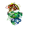 8b4vC  8b4wC 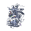 8oyhC  5jxhS S: Starting model for refinement C: citing same article ( |
|---|---|
| Similar structure data |
-Links
 PDBj
PDBj


- Assembly
| Deposited unit | 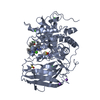
| ||||||||||||
|---|---|---|---|---|---|---|---|---|---|---|---|---|---|
| 1 |
| ||||||||||||
| Unit cell |
| ||||||||||||
| Components on special symmetry positions |
|
-Components
-Protein / Protein/peptide , 2 types, 2 molecules AB
| #1: Protein | Mass: 52112.312 Da / Num. of mol.: 1 Source method: isolated from a genetically manipulated source Source: (gene. exp.) Homo sapiens (human) / Gene: FURIN, FUR, PACE, PCSK3 / Cell line (production host): HEK293S / Production host: Homo sapiens (human) / References: UniProt: P09958, furin |
|---|---|
| #2: Protein/peptide | Mass: 750.957 Da / Num. of mol.: 1 / Source method: obtained synthetically / Source: (synth.) synthetic construct (others) |
-Non-polymers , 5 types, 465 molecules 


| #3: Chemical |  Mass: 40.078 Da / Num. of mol.: 3 / Source method: obtained synthetically / Formula: Ca Mass: 40.078 Da / Num. of mol.: 3 / Source method: obtained synthetically / Formula: Ca#4: Chemical | ChemComp-NA /  Mass: 22.990 Da / Num. of mol.: 4 / Source method: obtained synthetically / Formula: Na Mass: 22.990 Da / Num. of mol.: 4 / Source method: obtained synthetically / Formula: Na#5: Chemical | ChemComp-CL / |  Mass: 35.453 Da / Num. of mol.: 1 / Source method: obtained synthetically / Formula: Cl Mass: 35.453 Da / Num. of mol.: 1 / Source method: obtained synthetically / Formula: Cl#6: Chemical |  Mass: 78.133 Da / Num. of mol.: 2 / Source method: obtained synthetically / Formula: C2H6OS / Comment: DMSO, precipitant*YM Mass: 78.133 Da / Num. of mol.: 2 / Source method: obtained synthetically / Formula: C2H6OS / Comment: DMSO, precipitant*YM#7: Water | ChemComp-HOH / | Mass: 18.015 Da / Num. of mol.: 455 / Source method: isolated from a natural source / Formula: H2O |
|---|
-Details
| Has ligand of interest | Y |
|---|
-Experimental details
-Experiment
| Experiment | Method: X-RAY DIFFRACTION / Number of used crystals: 1 |
|---|
- Sample preparation
| Crystal | Density Matthews: 3.75 Å3/Da / Density % sol: 67.24 % |
|---|---|
| Crystal grow | Temperature: 293.15 K / Method: vapor diffusion, sitting drop Details: CRYSTALLIZATION SOLUTION: 100mM MES, 200mM K/NAH2PO4, PH 5.5, 2 M NACL; RESERVOIR SOLUTION: 3.0-3.2M NACL |
-Data collection
| Diffraction | Mean temperature: 100 K / Serial crystal experiment: N |
|---|---|
| Diffraction source | Source: SYNCHROTRON / Site: ESRF  / Beamline: ID30B / Wavelength: 0.968626 Å / Beamline: ID30B / Wavelength: 0.968626 Å |
| Detector | Type: DECTRIS PILATUS3 6M / Detector: PIXEL / Date: Sep 27, 2020 |
| Radiation | Protocol: SINGLE WAVELENGTH / Monochromatic (M) / Laue (L): M / Scattering type: x-ray |
| Radiation wavelength | Wavelength: 0.968626 Å / Relative weight: 1 |
| Reflection | Resolution: 1.6→46.06 Å / Num. obs: 104100 / % possible obs: 99 % / Redundancy: 19 % / Biso Wilson estimate: 23.44 Å2 / CC1/2: 0.999 / Rrim(I) all: 0.153 / Net I/σ(I): 13.81 |
| Reflection shell | Resolution: 1.6→1.7 Å / Mean I/σ(I) obs: 1.22 / Num. unique obs: 16441 / CC1/2: 0.513 / Rrim(I) all: 3.276 / % possible all: 98.1 |
- Processing
| Software |
| |||||||||||||||||||||||||||||||||||||||||||||||||||||||||||||||||||||||||||||||||||||||||||||||||||||||||||||||||||||||||||||||||||||||||||||||||||||||||||||||||||||||||||||||||||||||||||||||||||||||||||||||||||||||||
|---|---|---|---|---|---|---|---|---|---|---|---|---|---|---|---|---|---|---|---|---|---|---|---|---|---|---|---|---|---|---|---|---|---|---|---|---|---|---|---|---|---|---|---|---|---|---|---|---|---|---|---|---|---|---|---|---|---|---|---|---|---|---|---|---|---|---|---|---|---|---|---|---|---|---|---|---|---|---|---|---|---|---|---|---|---|---|---|---|---|---|---|---|---|---|---|---|---|---|---|---|---|---|---|---|---|---|---|---|---|---|---|---|---|---|---|---|---|---|---|---|---|---|---|---|---|---|---|---|---|---|---|---|---|---|---|---|---|---|---|---|---|---|---|---|---|---|---|---|---|---|---|---|---|---|---|---|---|---|---|---|---|---|---|---|---|---|---|---|---|---|---|---|---|---|---|---|---|---|---|---|---|---|---|---|---|---|---|---|---|---|---|---|---|---|---|---|---|---|---|---|---|---|---|---|---|---|---|---|---|---|---|---|---|---|---|---|---|---|
| Refinement | Method to determine structure: MOLECULAR REPLACEMENT Starting model: 5jxh Resolution: 1.6→46.06 Å / SU ML: 0.1922 / Cross valid method: FREE R-VALUE / σ(F): 1.35 / Phase error: 17.7426 Stereochemistry target values: GeoStd + Monomer Library + CDL v1.2
| |||||||||||||||||||||||||||||||||||||||||||||||||||||||||||||||||||||||||||||||||||||||||||||||||||||||||||||||||||||||||||||||||||||||||||||||||||||||||||||||||||||||||||||||||||||||||||||||||||||||||||||||||||||||||
| Solvent computation | Shrinkage radii: 0.9 Å / VDW probe radii: 1.1 Å / Solvent model: FLAT BULK SOLVENT MODEL | |||||||||||||||||||||||||||||||||||||||||||||||||||||||||||||||||||||||||||||||||||||||||||||||||||||||||||||||||||||||||||||||||||||||||||||||||||||||||||||||||||||||||||||||||||||||||||||||||||||||||||||||||||||||||
| Displacement parameters | Biso mean: 28.95 Å2 | |||||||||||||||||||||||||||||||||||||||||||||||||||||||||||||||||||||||||||||||||||||||||||||||||||||||||||||||||||||||||||||||||||||||||||||||||||||||||||||||||||||||||||||||||||||||||||||||||||||||||||||||||||||||||
| Refinement step | Cycle: LAST / Resolution: 1.6→46.06 Å
| |||||||||||||||||||||||||||||||||||||||||||||||||||||||||||||||||||||||||||||||||||||||||||||||||||||||||||||||||||||||||||||||||||||||||||||||||||||||||||||||||||||||||||||||||||||||||||||||||||||||||||||||||||||||||
| Refine LS restraints |
| |||||||||||||||||||||||||||||||||||||||||||||||||||||||||||||||||||||||||||||||||||||||||||||||||||||||||||||||||||||||||||||||||||||||||||||||||||||||||||||||||||||||||||||||||||||||||||||||||||||||||||||||||||||||||
| LS refinement shell |
| |||||||||||||||||||||||||||||||||||||||||||||||||||||||||||||||||||||||||||||||||||||||||||||||||||||||||||||||||||||||||||||||||||||||||||||||||||||||||||||||||||||||||||||||||||||||||||||||||||||||||||||||||||||||||
| Refinement TLS params. | Method: refined / Origin x: 35.3403029172 Å / Origin y: -37.8053366253 Å / Origin z: 0.128744691874 Å
| |||||||||||||||||||||||||||||||||||||||||||||||||||||||||||||||||||||||||||||||||||||||||||||||||||||||||||||||||||||||||||||||||||||||||||||||||||||||||||||||||||||||||||||||||||||||||||||||||||||||||||||||||||||||||
| Refinement TLS group | Selection details: chain A |
