[English] 日本語
Yorodumi
- PDB-8b3s: Structure of YjbA in complex with ClpC N-terminal Domain -

+
Open data


ID or keywords:

Loading...

-
Basic information

Entry
Database: PDB / ID: 8b3s
TitleStructure of YjbA in complex with ClpC N-terminal Domain
Components
  • ATP-dependent Clp protease ATP-binding subunit ClpC / Negative regulator of tic competence clcC/mecB
  • UPF0736 protein B4122_0676,UPF0736 protein YjbA
KeywordsUNKNOWN FUNCTION / sporulation
Function / homology
Function and homology information


peptidase activity / ATP hydrolysis activity / proteolysis / ATP binding
Similarity search - Function
UPF0736 / Protein of unknown function (DUF3603) / UVR domain / UVR domain profile. / ClpA/B, conserved site 2 / Chaperonins clpA/B signature 2. / ClpA/B, conserved site 1 / Chaperonins clpA/B signature 1. / ClpA/ClpB, AAA lid domain / AAA lid domain ...UPF0736 / Protein of unknown function (DUF3603) / UVR domain / UVR domain profile. / ClpA/B, conserved site 2 / Chaperonins clpA/B signature 2. / ClpA/B, conserved site 1 / Chaperonins clpA/B signature 1. / ClpA/ClpB, AAA lid domain / AAA lid domain / Clp amino terminal domain, pathogenicity island component / Clp repeat (R) domain profile. / Clp, repeat (R) domain / Clp, N-terminal domain superfamily / ClpA/B family / Clp ATPase, C-terminal / AAA domain (Cdc48 subfamily) / C-terminal, D2-small domain, of ClpB protein / C-terminal, D2-small domain, of ClpB protein / ATPase family associated with various cellular activities (AAA) / ATPase, AAA-type, core / ATPases associated with a variety of cellular activities / AAA+ ATPase domain / P-loop containing nucleoside triphosphate hydrolase
Similarity search - Domain/homology
UPF0736 protein B4122_0676 / ATP-dependent Clp protease ATP-binding subunit ClpC / Negative regulator of tic competence clcC/mecB / UPF0736 protein YjbA
Similarity search - Component
Biological speciesBacillus subtilis (bacteria)
MethodX-RAY DIFFRACTION / SYNCHROTRON / MOLECULAR REPLACEMENT / Resolution: 2.09 Å
AuthorsEvans, N.J. / Isaacson, R.L. / Camp, A.H. / Collins, M.K.
Funding support United Kingdom, 1items
OrganizationGrant numberCountry
Biotechnology and Biological Sciences Research Council (BBSRC) United Kingdom
CitationJournal: To Be Published
Title: Structure of YjbA in complex with ClpC N-terminal Domain
Authors: Evans, N.J. / Isaacson, R.L.
History
DepositionSep 16, 2022Deposition site: PDBE / Processing site: PDBE
Revision 1.0Dec 27, 2023Provider: repository / Type: Initial release

-
Structure visualization

Structure viewerMolecule:
MolmilJmol/JSmol

Downloads & links

-
Assembly

Deposited unit
A: UPF0736 protein B4122_0676,UPF0736 protein YjbA
Z: ATP-dependent Clp protease ATP-binding subunit ClpC / Negative regulator of tic competence clcC/mecB
hetero molecules


Theoretical massNumber of molelcules
Total (without water)59,8959
Polymers59,3182
Non-polymers5777
Water4,360242
1


  • Idetical with deposited unit
  • defined by author
  • Evidence: gel filtration, Peak shifting was observed upon binding, isothermal titration calorimetry, Fitted data for YjbA ClpC interaction: dH = -19.5 +/- 0.6 kJ/mol; KD = 3.79 +/- 0.50 uM; N = 0.82 ...Evidence: gel filtration, Peak shifting was observed upon binding, isothermal titration calorimetry, Fitted data for YjbA ClpC interaction: dH = -19.5 +/- 0.6 kJ/mol; KD = 3.79 +/- 0.50 uM; N = 0.82 +/- 0.01 sites., assay for oligomerization, In an NMR Titration observing chemical shift perturbation peaks disappeared upon binding in a dose dependant manner.
TypeNameSymmetry operationNumber
identity operation1_555x,y,z1
Unit cell
Length a, b, c (Å)69.713, 69.713, 89.601
Angle α, β, γ (deg.)90.00, 90.00, 120.00
Int Tables number144
Space group name H-MP31

-
Components

#1: Protein UPF0736 protein B4122_0676,UPF0736 protein YjbA


Mass: 42370.293 Da / Num. of mol.: 1
Source method: isolated from a genetically manipulated source
Source: (gene. exp.) Bacillus subtilis (bacteria)
Gene: B4122_0676, B4417_4067, Bateq7PJ16_1268, DFO69_2911, J5227_16610, SC09_Contig19orf00439, yjbA, BSU11410
Strain: 168 / Production host: Escherichia coli (E. coli) / References: UniProt: A0A085CA92, UniProt: O31597
#2: Protein ATP-dependent Clp protease ATP-binding subunit ClpC / Negative regulator of tic competence clcC/mecB


Mass: 16947.436 Da / Num. of mol.: 1
Source method: isolated from a genetically manipulated source
Source: (gene. exp.) Bacillus subtilis (bacteria) / Gene: B4417_1475 / Production host: Escherichia coli (E. coli) / References: UniProt: A0A164W157
#3: Chemical
ChemComp-GOL / GLYCEROL / GLYCERIN / PROPANE-1,2,3-TRIOL / Glycerol


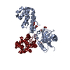
Mass: 92.094 Da / Num. of mol.: 6 / Source method: obtained synthetically / Formula: C3H8O3
#4: Chemical ChemComp-MG / MAGNESIUM ION


Mass: 24.305 Da / Num. of mol.: 1 / Source method: obtained synthetically / Formula: Mg
#5: Water ChemComp-HOH / water / Water


Mass: 18.015 Da / Num. of mol.: 242 / Source method: isolated from a natural source / Formula: H2O
Has ligand of interestN

-
Experimental details

-
Experiment

ExperimentMethod: X-RAY DIFFRACTION / Number of used crystals: 1

-
Sample preparation

CrystalDensity Matthews: 2.12 Å3/Da / Density % sol: 41.96 % / Description: Rods
Crystal growTemperature: 289 K / Method: vapor diffusion, sitting drop / pH: 7 / Details: 4% PEG 8000, 0.2 M Magnesium Chloride / Temp details: bench top incubator

-
Data collection

DiffractionMean temperature: 100 K / Serial crystal experiment: N
Diffraction sourceSource: SYNCHROTRON / Site: Diamond / Beamline: I03 / Wavelength: 0.9763 Å
DetectorType: DECTRIS EIGER2 S 16M / Detector: PIXEL / Date: Sep 17, 2017
RadiationProtocol: SINGLE WAVELENGTH / Monochromatic (M) / Laue (L): M / Scattering type: x-ray
Radiation wavelengthWavelength: 0.9763 Å / Relative weight: 1
ReflectionResolution: 2.09→29.9 Å / Num. obs: 28876 / % possible obs: 100 % / Redundancy: 8.6 % / CC1/2: 1 / Net I/σ(I): 9.8
Reflection shellResolution: 2.09→2.13 Å / Redundancy: 8.8 % / Num. unique obs: 1440 / CC1/2: 0.5 / % possible all: 100

-
Processing

Software
NameVersionClassification
PHENIX(1.16_3549: ???)refinement
DIALSdata reduction
DIALSdata scaling
PHASERphasing
SHELXDEphasing
ARP/wARPmodel building
BUCCANEERmodel building
RefinementMethod to determine structure: MOLECULAR REPLACEMENT
Starting model: 5hbn
Resolution: 2.09→29.867 Å / SU ML: 0.36 / Cross valid method: FREE R-VALUE / σ(F): 1.96 / Phase error: 28.11 / Stereochemistry target values: ML
RfactorNum. reflection% reflection
Rfree0.234 1470 5.11 %
Rwork0.1846 --
obs0.1871 28744 99.67 %
Solvent computationShrinkage radii: 0.9 Å / VDW probe radii: 1.11 Å / Solvent model: FLAT BULK SOLVENT MODEL
Refinement stepCycle: LAST / Resolution: 2.09→29.867 Å
ProteinNucleic acidLigandSolventTotal
Num. atoms3114 0 37 242 3393
Refine LS restraints
Refine-IDTypeDev idealNumber
X-RAY DIFFRACTIONf_bond_d0.0023249
X-RAY DIFFRACTIONf_angle_d0.5144387
X-RAY DIFFRACTIONf_dihedral_angle_d4.2872686
X-RAY DIFFRACTIONf_chiral_restr0.037475
X-RAY DIFFRACTIONf_plane_restr0.004562
LS refinement shell
Resolution (Å)Rfactor RfreeNum. reflection RfreeRfactor RworkNum. reflection RworkRefine-ID% reflection obs (%)
2.09-2.16450.43091510.33852696X-RAY DIFFRACTION99
2.1645-2.25120.33331670.29632725X-RAY DIFFRACTION100
2.2512-2.35360.34221490.2762708X-RAY DIFFRACTION100
2.3536-2.47760.3061210.23962750X-RAY DIFFRACTION100
2.4776-2.63280.28511340.2152753X-RAY DIFFRACTION100
2.6328-2.83590.26851540.20562714X-RAY DIFFRACTION100
2.8359-3.1210.23691760.18342695X-RAY DIFFRACTION100
3.121-3.5720.21861200.16042755X-RAY DIFFRACTION100
3.572-4.49790.18031540.13072733X-RAY DIFFRACTION100
4.4979-29.8670.17411440.15282745X-RAY DIFFRACTION100
Refinement TLS params.Method: refined / Origin x: 16.0773 Å / Origin y: -0.9208 Å / Origin z: 2.6425 Å
111213212223313233
T0.142 Å2-0.0195 Å2-0.0037 Å2-0.3377 Å2-0.0311 Å2--0.2353 Å2
L0.443 °2-0.182 °2-0.146 °2-1.2259 °20.1075 °2--1.7687 °2
S0.0061 Å °0.0972 Å °-0.0191 Å °-0.0484 Å °0.0626 Å °-0.0724 Å °-0.0329 Å °0.355 Å °-0.0505 Å °
Refinement TLS groupSelection details: all

+
About Yorodumi

-
News

-
Feb 9, 2022. New format data for meta-information of EMDB entries

New format data for meta-information of EMDB entries

  • Version 3 of the EMDB header file is now the official format.
  • The previous official version 1.9 will be removed from the archive.

Related info.:EMDB header

External links:wwPDB to switch to version 3 of the EMDB data model

-
Aug 12, 2020. Covid-19 info

Covid-19 info

URL: https://pdbj.org/emnavi/covid19.php

New page: Covid-19 featured information page in EM Navigator.

Related info.:Covid-19 info / Mar 5, 2020. Novel coronavirus structure data

+
Mar 5, 2020. Novel coronavirus structure data

Novel coronavirus structure data

Related info.:Yorodumi Speices / Aug 12, 2020. Covid-19 info

External links:COVID-19 featured content - PDBj / Molecule of the Month (242):Coronavirus Proteases

+
Jan 31, 2019. EMDB accession codes are about to change! (news from PDBe EMDB page)

EMDB accession codes are about to change! (news from PDBe EMDB page)

  • The allocation of 4 digits for EMDB accession codes will soon come to an end. Whilst these codes will remain in use, new EMDB accession codes will include an additional digit and will expand incrementally as the available range of codes is exhausted. The current 4-digit format prefixed with “EMD-” (i.e. EMD-XXXX) will advance to a 5-digit format (i.e. EMD-XXXXX), and so on. It is currently estimated that the 4-digit codes will be depleted around Spring 2019, at which point the 5-digit format will come into force.
  • The EM Navigator/Yorodumi systems omit the EMD- prefix.

Related info.:Q: What is EMD? / ID/Accession-code notation in Yorodumi/EM Navigator

External links:EMDB Accession Codes are Changing Soon! / Contact to PDBj

+
Jul 12, 2017. Major update of PDB

Major update of PDB

  • wwPDB released updated PDB data conforming to the new PDBx/mmCIF dictionary.
  • This is a major update changing the version number from 4 to 5, and with Remediation, in which all the entries are updated.
  • In this update, many items about electron microscopy experimental information are reorganized (e.g. em_software).
  • Now, EM Navigator and Yorodumi are based on the updated data.

External links:wwPDB Remediation / Enriched Model Files Conforming to OneDep Data Standards Now Available in the PDB FTP Archive

-
Yorodumi

Thousand views of thousand structures

  • Yorodumi is a browser for structure data from EMDB, PDB, SASBDB, etc.
  • This page is also the successor to EM Navigator detail page, and also detail information page/front-end page for Omokage search.
  • The word "yorodu" (or yorozu) is an old Japanese word meaning "ten thousand". "mi" (miru) is to see.

Related info.:EMDB / PDB / SASBDB / Comparison of 3 databanks / Yorodumi Search / Aug 31, 2016. New EM Navigator & Yorodumi / Yorodumi Papers / Jmol/JSmol / Function and homology information / Changes in new EM Navigator and Yorodumi

Read more