 Movie
Movie Controller

[English] 日本語
 Yorodumi
- PDB-8b38: Chaetoceros socialis forma radians RNA virus 1 full capsid atomic... -
+ Open data

- Basic information
| Entry | Database: PDB / ID: 8b38 | ||||||
|---|---|---|---|---|---|---|---|
| Title | Chaetoceros socialis forma radians RNA virus 1 full capsid atomic model | ||||||
 Components Components | (Structural polyprotein) x 3 | ||||||
 Keywords Keywords | VIRUS / pseudo-T=3 / icosahedral | ||||||
| Function / homology |  Function and homology information Function and homology information | ||||||
| Biological species |  Chaetoceros socialis forma radians RNA virus 1 Chaetoceros socialis forma radians RNA virus 1 | ||||||
| Method | ELECTRON MICROSCOPY / single particle reconstruction / cryo EM / Resolution: 3 Å | ||||||
 Authors Authors | Wang, H. / Okamoto, K. / Munke, A. | ||||||
| Funding support |  Sweden, 1items Sweden, 1items
| ||||||
 Citation Citation | Journal: Viruses / Year: 2022 Title: Structural Insights into Common and Host-Specific Receptor-Binding Mechanisms in Algal Picorna-like Viruses. Authors: Han Wang / Anna Munke / Siqi Li / Yuji Tomaru / Kenta Okamoto /   Abstract: viruses are abundant algal viruses that regulate the dynamics of algal blooms in aquatic environments. They employ a narrow host range because they merely lyse their algal host species. This host- ... viruses are abundant algal viruses that regulate the dynamics of algal blooms in aquatic environments. They employ a narrow host range because they merely lyse their algal host species. This host-specific lysis is thought to correspond to the unique receptor-binding mechanism of the viruses. Here, we present the atomic structures of the full and empty capsids of Chaetoceros socialis forma radians RNA virus 1 built-in 3.0 Å and 3.1 Å cryo-electron microscopy maps. The empty capsid structure and the structural variability provide insights into its assembly and uncoating intermediates. In conjunction with the previously reported atomic model of the Chaetoceros tenuissimus RNA virus type II capsid, we have identified the common and diverse structural features of the VP1 surface between the viruses. We have also tested the potential usage of AlphaFold2 for structural prediction of the VP1s and a subsequent structural phylogeny for classifying viruses by their hosts. These findings will be crucial for inferring the host-specific receptor-binding mechanism in viruses. | ||||||
| History |
|
- Structure visualization
| Structure viewer | Molecule: MolmilJmol/JSmol |
|---|
- Downloads & links
-Download
| PDBx/mmCIF format | 8b38.cif.gz | 172.4 KB | Display | PDBx/mmCIF format |
|---|---|---|---|---|
| PDB format | pdb8b38.ent.gz | 131.6 KB | Display | PDB format |
| PDBx/mmJSON format | 8b38.json.gz | Tree view | PDBx/mmJSON format | |
| Others |  Other downloads Other downloads |
-Validation report
| Arichive directory | https://data.pdbj.org/pub/pdb/validation_reports/b3/8b38ftp://data.pdbj.org/pub/pdb/validation_reports/b3/8b38 | HTTPS FTP |
|---|
-Related structure data
| Related structure data |  15823MC  8b3jC M: map data used to model this data C: citing same article ( |
|---|---|
| Similar structure data |

-Links
 PDBj
PDBj- Assembly
| Deposited unit | 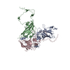
|
|---|---|
| 1 | x 60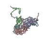
|
-Components
| #1: Protein | Mass: 29930.619 Da / Num. of mol.: 1 / Source method: isolated from a natural source Source: (natural) Chaetoceros socialis forma radians RNA virus 1References: UniProt: B9A8E1 |
|---|---|
| #2: Protein | Mass: 26871.293 Da / Num. of mol.: 1 / Source method: isolated from a natural source Source: (natural) Chaetoceros socialis forma radians RNA virus 1References: UniProt: B9A8E1 |
| #3: Protein | Mass: 31098.668 Da / Num. of mol.: 1 / Source method: isolated from a natural source Source: (natural) Chaetoceros socialis forma radians RNA virus 1References: UniProt: B9A8E1 |
-Experimental details
-Experiment
| Experiment | Method: ELECTRON MICROSCOPY |
|---|---|
| EM experiment | Aggregation state: PARTICLE / 3D reconstruction method: single particle reconstruction |
- Sample preparation
| Component | Name: Chaetoceros socialis forma radians RNA virus 1 / Type: VIRUS / Entity ID: all / Source: NATURAL |
|---|---|
| Source (natural) | Organism: Chaetoceros socialis forma radians RNA virus 1 |
| Details of virus | Empty: NO / Enveloped: NO / Isolate: STRAIN / Type: VIRION |
| Buffer solution | pH: 7 |
| Specimen | Embedding applied: NO / Shadowing applied: NO / Staining applied: NO / Vitrification applied: YES |
| Vitrification | Cryogen name: ETHANE |
- Electron microscopy imaging
| Experimental equipment |  Model: Titan Krios / Image courtesy: FEI Company |
|---|---|
| Microscopy | Model: FEI TITAN KRIOS |
| Electron gun | Electron source:  FIELD EMISSION GUN / Accelerating voltage: 300 kV / Illumination mode: SPOT SCAN FIELD EMISSION GUN / Accelerating voltage: 300 kV / Illumination mode: SPOT SCAN |
| Electron lens | Mode: BRIGHT FIELD / Nominal defocus max: 4000 nm / Nominal defocus min: 1000 nm |
| Image recording | Electron dose: 30 e/Å2 / Film or detector model: GATAN K3 (6k x 4k) |
- Processing
| CTF correction | Type: PHASE FLIPPING ONLY |
|---|---|
| 3D reconstruction | Resolution: 3 Å / Resolution method: FSC 0.143 CUT-OFF / Num. of particles: 3984 / Symmetry type: POINT |
