 Movie
Movie Controller


+ Open data

- Basic information
| Entry | Database: PDB / ID: 8b2p | ||||||||||||
|---|---|---|---|---|---|---|---|---|---|---|---|---|---|
| Title | CYP153A71 from Acinetobacter dieselolei bound to octanoic acid | ||||||||||||
 Components Components | Cytochrome P450 alkane hydroxylase | ||||||||||||
 Keywords Keywords | OXIDOREDUCTASE / Alkane hydroxylase Cytochrome P450 monooxygenase CYP153 Octanoic acid | ||||||||||||
| Function / homology |  Function and homology information Function and homology informationoxidoreductase activity, acting on paired donors, with incorporation or reduction of molecular oxygen / monooxygenase activity / iron ion binding / heme binding Similarity search - Function | ||||||||||||
| Biological species |  Alcanivorax dieselolei (bacteria) Alcanivorax dieselolei (bacteria) | ||||||||||||
| Method |  X-RAY DIFFRACTION / SYNCHROTRON / MOLECULAR REPLACEMENT / molecular replacement / Resolution: 1.95 Å X-RAY DIFFRACTION / SYNCHROTRON / MOLECULAR REPLACEMENT / molecular replacement / Resolution: 1.95 Å | ||||||||||||
 Authors Authors | Opperman, D.J. / Tolmie, C. | ||||||||||||
| Funding support |  United Kingdom, United Kingdom,  South Africa, 3items South Africa, 3items
| ||||||||||||
 Citation Citation | Journal: Catalysts / Year: 2022 Title: CYP153A71 from Alcanivorax dieselolei: Oxidation beyond Monoterminal Hydroxylation of n-Alkanes Authors: Jacobs, C.L. / do Aido-Machado, R. / Tolmie, C. / Smit, M.S. / Opperman, D.J. | ||||||||||||
| History |
|
- Structure visualization
| Structure viewer | Molecule: MolmilJmol/JSmol |
|---|
- Downloads & links
-Download
| PDBx/mmCIF format | 8b2p.cif.gz | 192.5 KB | Display | PDBx/mmCIF format |
|---|---|---|---|---|
| PDB format | pdb8b2p.ent.gz | 151 KB | Display | PDB format |
| PDBx/mmJSON format | 8b2p.json.gz | Tree view | PDBx/mmJSON format | |
| Others |  Other downloads Other downloads |
-Validation report
| Arichive directory | https://data.pdbj.org/pub/pdb/validation_reports/b2/8b2pftp://data.pdbj.org/pub/pdb/validation_reports/b2/8b2p | HTTPS FTP |
|---|
-Related structure data
| Related structure data |  5fyfS S: Starting model for refinement |
|---|---|
| Similar structure data |
-Links
 PDBj
PDBj
- Assembly
| Deposited unit | 
| ||||||||||||||||||
|---|---|---|---|---|---|---|---|---|---|---|---|---|---|---|---|---|---|---|---|
| 1 | 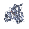
| ||||||||||||||||||
| 2 | 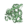
| ||||||||||||||||||
| Unit cell |
| ||||||||||||||||||
| Noncrystallographic symmetry (NCS) | NCS domain:
NCS domain segments: Component-ID: _ / Ens-ID: 1 / Beg auth comp-ID: GLN / Beg label comp-ID: GLN / End auth comp-ID: ALA / End label comp-ID: ALA / Refine code: _ / Auth seq-ID: 49 - 468 / Label seq-ID: 49 - 468
|
-Components
| #1: Protein | Mass: 53881.699 Da / Num. of mol.: 2 Source method: isolated from a genetically manipulated source Source: (gene. exp.) Alcanivorax dieselolei (bacteria) / Gene: p450 / Production host: #2: Chemical |   Mass: 616.487 Da / Num. of mol.: 2 / Source method: obtained synthetically / Formula: C34H32FeN4O4 / Feature type: SUBJECT OF INVESTIGATION Mass: 616.487 Da / Num. of mol.: 2 / Source method: obtained synthetically / Formula: C34H32FeN4O4 / Feature type: SUBJECT OF INVESTIGATION#3: Chemical | 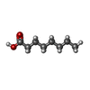  Mass: 144.211 Da / Num. of mol.: 2 / Source method: obtained synthetically / Formula: C8H16O2 Mass: 144.211 Da / Num. of mol.: 2 / Source method: obtained synthetically / Formula: C8H16O2#4: Water | ChemComp-HOH / |  Mass: 18.015 Da / Num. of mol.: 281 / Source method: isolated from a natural source / Formula: H2O Mass: 18.015 Da / Num. of mol.: 281 / Source method: isolated from a natural source / Formula: H2OHas ligand of interest | Y | |
|---|
-Experimental details
-Experiment
| Experiment | Method: X-RAY DIFFRACTION / Number of used crystals: 1 |
|---|
- Sample preparation
| Crystal | Density Matthews: 2.21 Å3/Da / Density % sol: 44.24 % |
|---|---|
| Crystal grow | Temperature: 289 K / Method: vapor diffusion / pH: 7.5 Details: 0.1 M HEPES pH 7.5 12% (w/v) polyethylene glycol 3350 10 mM octanoic acid |
-Data collection
| Diffraction | Mean temperature: 100 K / Serial crystal experiment: N | ||||||||||||||||||||||||||||||
|---|---|---|---|---|---|---|---|---|---|---|---|---|---|---|---|---|---|---|---|---|---|---|---|---|---|---|---|---|---|---|---|
| Diffraction source | Source: SYNCHROTRON / Site: Diamond / Beamline: I03 / Wavelength: 0.9763 Å | ||||||||||||||||||||||||||||||
| Detector | Type: DECTRIS PILATUS3 6M / Detector: PIXEL / Date: Oct 14, 2018 | ||||||||||||||||||||||||||||||
| Radiation | Protocol: SINGLE WAVELENGTH / Monochromatic (M) / Laue (L): M / Scattering type: x-ray | ||||||||||||||||||||||||||||||
| Radiation wavelength | Wavelength: 0.9763 Å / Relative weight: 1 | ||||||||||||||||||||||||||||||
| Reflection | Resolution: 1.95→46.11 Å / Num. obs: 66934 / % possible obs: 97.8 % / Redundancy: 3.4 % / CC1/2: 0.995 / Rmerge(I) obs: 0.129 / Rpim(I) all: 0.083 / Rrim(I) all: 0.154 / Net I/σ(I): 7.2 / Num. measured all: 227101 / Scaling rejects: 23 | ||||||||||||||||||||||||||||||
| Reflection shell | Diffraction-ID: 1
|
-Phasing
| Phasing | Method: molecular replacement | |||||||||
|---|---|---|---|---|---|---|---|---|---|---|
| Phasing MR | Model details: Phaser MODE: MR_AUTO
|
- Processing
| Software |
| ||||||||||||||||||||||||||||||||||||||||||||||||||||||||||||
|---|---|---|---|---|---|---|---|---|---|---|---|---|---|---|---|---|---|---|---|---|---|---|---|---|---|---|---|---|---|---|---|---|---|---|---|---|---|---|---|---|---|---|---|---|---|---|---|---|---|---|---|---|---|---|---|---|---|---|---|---|---|
| Refinement | Method to determine structure: MOLECULAR REPLACEMENT Starting model: 5FYF Resolution: 1.95→46.11 Å / Cor.coef. Fo:Fc: 0.952 / Cor.coef. Fo:Fc free: 0.929 / WRfactor Rfree: 0.2547 / WRfactor Rwork: 0.2119 / FOM work R set: 0.5859 / SU B: 11.558 / SU ML: 0.277 / SU R Cruickshank DPI: 0.2104 / SU Rfree: 0.1831 / Cross valid method: THROUGHOUT / σ(F): 0 / ESU R: 0.21 / ESU R Free: 0.183 / Stereochemistry target values: MAXIMUM LIKELIHOOD Details: HYDROGENS HAVE BEEN ADDED IN THE RIDING POSITIONS U VALUES : REFINED INDIVIDUALLY
| ||||||||||||||||||||||||||||||||||||||||||||||||||||||||||||
| Solvent computation | Ion probe radii: 0.8 Å / Shrinkage radii: 0.8 Å / VDW probe radii: 1.2 Å / Solvent model: MASK | ||||||||||||||||||||||||||||||||||||||||||||||||||||||||||||
| Displacement parameters | Biso max: 137.25 Å2 / Biso mean: 40.926 Å2 / Biso min: 14.72 Å2
| ||||||||||||||||||||||||||||||||||||||||||||||||||||||||||||
| Refinement step | Cycle: final / Resolution: 1.95→46.11 Å
| ||||||||||||||||||||||||||||||||||||||||||||||||||||||||||||
| Refine LS restraints |
| ||||||||||||||||||||||||||||||||||||||||||||||||||||||||||||
| Refine LS restraints NCS | Ens-ID: 1 / Number: 14481 / Refine-ID: X-RAY DIFFRACTION / Type: interatomic distance / Rms dev position: 0.04 Å / Weight position: 0.05
| ||||||||||||||||||||||||||||||||||||||||||||||||||||||||||||
| LS refinement shell | Resolution: 1.95→2.001 Å / Rfactor Rfree error: 0 / Total num. of bins used: 20
|
