 Movie
Movie Controller

[English] 日本語
 Yorodumi
- PDB-8b2o: Millisecond cryo-trapping by the spitrobot crystal plunger, CTX-M... -
+ Open data

- Basic information
| Entry | Database: PDB / ID: 8b2o | ||||||
|---|---|---|---|---|---|---|---|
| Title | Millisecond cryo-trapping by the spitrobot crystal plunger, CTX-M-14 E166A, Ampicillin, 5 sec | ||||||
 Components Components | Beta-lactamase | ||||||
 Keywords Keywords | HYDROLASE / beta-lactamase / CTX-M-14 / time-resolved crystallography | ||||||
| Function / homology |  Function and homology information Function and homology informationbeta-lactam antibiotic catabolic process / beta-lactamase activity / beta-lactamase / response to antibiotic Similarity search - Function | ||||||
| Biological species |  Klebsiella pneumoniae (bacteria) Klebsiella pneumoniae (bacteria) | ||||||
| Method |  X-RAY DIFFRACTION / SYNCHROTRON / MOLECULAR REPLACEMENT / Resolution: 1.86 Å X-RAY DIFFRACTION / SYNCHROTRON / MOLECULAR REPLACEMENT / Resolution: 1.86 Å | ||||||
 Authors Authors | Mehrabi, P. / Sung, S. / von Stetten, D. / Prester, A. / Hatton, C.E. / Kleine-Doepke, S. / Berkes, A. / Gore, G. / Leimkohl, J.P. / Schikora, H. ...Mehrabi, P. / Sung, S. / von Stetten, D. / Prester, A. / Hatton, C.E. / Kleine-Doepke, S. / Berkes, A. / Gore, G. / Leimkohl, J.P. / Schikora, H. / Kollewe, M. / Rohde, H. / Wilmanns, M. / Tellkamp, F. / Schulz, E.C. | ||||||
| Funding support |  Germany, 1items Germany, 1items
| ||||||
 Citation Citation | Journal: Nat Commun / Year: 2023 Title: Millisecond cryo-trapping by the spitrobot crystal plunger simplifies time-resolved crystallography. Authors: Mehrabi, P. / Sung, S. / von Stetten, D. / Prester, A. / Hatton, C.E. / Kleine-Dopke, S. / Berkes, A. / Gore, G. / Leimkohl, J.P. / Schikora, H. / Kollewe, M. / Rohde, H. / Wilmanns, M. / ...Authors: Mehrabi, P. / Sung, S. / von Stetten, D. / Prester, A. / Hatton, C.E. / Kleine-Dopke, S. / Berkes, A. / Gore, G. / Leimkohl, J.P. / Schikora, H. / Kollewe, M. / Rohde, H. / Wilmanns, M. / Tellkamp, F. / Schulz, E.C. | ||||||
| History |
|
- Structure visualization
| Structure viewer | Molecule: MolmilJmol/JSmol |
|---|
- Downloads & links
-Download
| PDBx/mmCIF format | 8b2o.cif.gz | 142.9 KB | Display | PDBx/mmCIF format |
|---|---|---|---|---|
| PDB format | pdb8b2o.ent.gz | 91.3 KB | Display | PDB format |
| PDBx/mmJSON format | 8b2o.json.gz | Tree view | PDBx/mmJSON format | |
| Others |  Other downloads Other downloads |
-Validation report
| Arichive directory | https://data.pdbj.org/pub/pdb/validation_reports/b2/8b2oftp://data.pdbj.org/pub/pdb/validation_reports/b2/8b2o | HTTPS FTP |
|---|
-Related structure data
| Related structure data |  8aw8C  8aw9C  8awbC  8awcC  8awdC 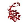 8aweC  8awfC 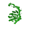 8awsC 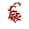 8awuC  8awvC 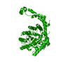 8awxC  8awyC  8b03C  8b05C  8b06C 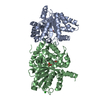 8b08C 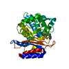 8b2vC 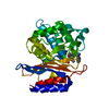 8b2wC  8b3mC 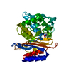 6gthS S: Starting model for refinement C: citing same article ( |
|---|---|
| Similar structure data |
-Links
 PDBj
PDBj


- Assembly
| Deposited unit | 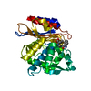
| ||||||||||||
|---|---|---|---|---|---|---|---|---|---|---|---|---|---|
| 1 |
| ||||||||||||
| Unit cell |
| ||||||||||||
| Components on special symmetry positions |
|
-Components
| #1: Protein | Mass: 27600.119 Da / Num. of mol.: 1 Source method: isolated from a genetically manipulated source Source: (gene. exp.) Klebsiella pneumoniae (bacteria) / Gene: ctx-m-14 / Production host: |
|---|---|
| #2: Chemical | ChemComp-AIX / (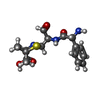  Mass: 351.421 Da / Num. of mol.: 1 / Source method: obtained synthetically / Formula: C16H21N3O4S / Feature type: SUBJECT OF INVESTIGATION Mass: 351.421 Da / Num. of mol.: 1 / Source method: obtained synthetically / Formula: C16H21N3O4S / Feature type: SUBJECT OF INVESTIGATION |
| #3: Water | ChemComp-HOH /  Mass: 18.015 Da / Num. of mol.: 146 / Source method: isolated from a natural source / Formula: H2O Mass: 18.015 Da / Num. of mol.: 146 / Source method: isolated from a natural source / Formula: H2O |
| Has ligand of interest | Y |
| Has protein modification | Y |
-Experimental details
-Experiment
| Experiment | Method: X-RAY DIFFRACTION / Number of used crystals: 1 |
|---|
- Sample preparation
| Crystal | Density Matthews: 2.13 Å3/Da / Density % sol: 42.25 % |
|---|---|
| Crystal grow | Temperature: 293 K / Method: batch mode / pH: 4.5 / Details: 40% PEG8000, 200 mM LiSO4, 100 mM NaOAc, pH 4.5 |
-Data collection
| Diffraction | Mean temperature: 100 K / Serial crystal experiment: N |
|---|---|
| Diffraction source | Source: SYNCHROTRON / Site: PETRA III, EMBL c/o DESY / Beamline: P14 (MX2) / Wavelength: 0.97626 Å |
| Detector | Type: DECTRIS EIGER2 X 16M / Detector: PIXEL / Date: Feb 22, 2022 |
| Radiation | Protocol: SINGLE WAVELENGTH / Monochromatic (M) / Laue (L): M / Scattering type: x-ray |
| Radiation wavelength | Wavelength: 0.97626 Å / Relative weight: 1 |
| Reflection | Resolution: 1.856→77.654 Å / Num. obs: 200593 / % possible obs: 0.807 % / Redundancy: 15.3 % / Biso Wilson estimate: 18.97 Å2 / CC1/2: 0.998 / Net I/σ(I): 8.8 |
| Reflection shell | Resolution: 1.856→2.094 Å / Num. unique obs: 10125 / CC1/2: 0.718 / % possible all: 37.2 |
- Processing
| Software |
| ||||||||||||||||||||||||||||||||||||||||||
|---|---|---|---|---|---|---|---|---|---|---|---|---|---|---|---|---|---|---|---|---|---|---|---|---|---|---|---|---|---|---|---|---|---|---|---|---|---|---|---|---|---|---|---|
| Refinement | Method to determine structure: MOLECULAR REPLACEMENT Starting model: 6gth Resolution: 1.86→77.65 Å / SU ML: 0.1954 / Cross valid method: FREE R-VALUE / σ(F): 1.36 / Phase error: 26.9746 Stereochemistry target values: GeoStd + Monomer Library + CDL v1.2
| ||||||||||||||||||||||||||||||||||||||||||
| Solvent computation | Shrinkage radii: 0.9 Å / VDW probe radii: 1.11 Å / Solvent model: FLAT BULK SOLVENT MODEL | ||||||||||||||||||||||||||||||||||||||||||
| Displacement parameters | Biso mean: 27.91 Å2 | ||||||||||||||||||||||||||||||||||||||||||
| Refinement step | Cycle: LAST / Resolution: 1.86→77.65 Å
| ||||||||||||||||||||||||||||||||||||||||||
| Refine LS restraints |
| ||||||||||||||||||||||||||||||||||||||||||
| LS refinement shell |
| ||||||||||||||||||||||||||||||||||||||||||
| Refinement TLS params. | Method: refined / Origin x: -4.13222624463 Å / Origin y: -4.32962394962 Å / Origin z: -19.0950026567 Å
| ||||||||||||||||||||||||||||||||||||||||||
| Refinement TLS group | Selection details: all |
