 Movie
Movie Controller


+ Open data

- Basic information
| Entry | Database: PDB / ID: 8b2h | ||||||
|---|---|---|---|---|---|---|---|
| Title | Muramidase from Thermothielavioides terrestris, catalytic domain | ||||||
 Components Components | SH3b domain-containing protein | ||||||
 Keywords Keywords | HYDROLASE / SH3-like / muramidase / peptidoglycan / cell wall binding domain | ||||||
| Function / homology | metal ion binding / Uncharacterized protein Function and homology information Function and homology information | ||||||
| Biological species |  Thermothielavioides terrestris (fungus) Thermothielavioides terrestris (fungus) | ||||||
| Method |  X-RAY DIFFRACTION / SYNCHROTRON / MOLECULAR REPLACEMENT / Resolution: 2.36 Å X-RAY DIFFRACTION / SYNCHROTRON / MOLECULAR REPLACEMENT / Resolution: 2.36 Å | ||||||
 Authors Authors | Moroz, O.V. / Blagova, E. / Lebedev, A.A. / Skov, L.K. / Pache, R.A. / Schnorr, K.M. / Kiemer, L. / Nymand-Grarup, S. / Ming, L. / Ye, L. ...Moroz, O.V. / Blagova, E. / Lebedev, A.A. / Skov, L.K. / Pache, R.A. / Schnorr, K.M. / Kiemer, L. / Nymand-Grarup, S. / Ming, L. / Ye, L. / Klausen, M. / Cohn, M.T. / Schmidt, E.G.W. / Davies, G.J. / Wilson, K.S. | ||||||
| Funding support | 1items
| ||||||
 Citation Citation | Journal: Acta Crystallogr D Struct Biol / Year: 2023 Title: Module walking using an SH3-like cell-wall-binding domain leads to a new GH184 family of muramidases. Authors: Moroz, O.V. / Blagova, E. / Lebedev, A.A. / Skov, L.K. / Pache, R.A. / Schnorr, K.M. / Kiemer, L. / Friis, E.P. / Nymand-Grarup, S. / Ming, L. / Ye, L. / Klausen, M. / Cohn, M.T. / Schmidt, ...Authors: Moroz, O.V. / Blagova, E. / Lebedev, A.A. / Skov, L.K. / Pache, R.A. / Schnorr, K.M. / Kiemer, L. / Friis, E.P. / Nymand-Grarup, S. / Ming, L. / Ye, L. / Klausen, M. / Cohn, M.T. / Schmidt, E.G.W. / Davies, G.J. / Wilson, K.S. | ||||||
| History |
|
- Structure visualization
| Structure viewer | Molecule: MolmilJmol/JSmol |
|---|
- Downloads & links
-Download
| PDBx/mmCIF format | 8b2h.cif.gz | 46.3 KB | Display | PDBx/mmCIF format |
|---|---|---|---|---|
| PDB format | pdb8b2h.ent.gz | 28.5 KB | Display | PDB format |
| PDBx/mmJSON format | 8b2h.json.gz | Tree view | PDBx/mmJSON format | |
| Others |  Other downloads Other downloads |
-Validation report
| Arichive directory | https://data.pdbj.org/pub/pdb/validation_reports/b2/8b2hftp://data.pdbj.org/pub/pdb/validation_reports/b2/8b2h | HTTPS FTP |
|---|
-Related structure data
| Related structure data |  8b2eSC  8b2fC  8b2gC  8b2sC S: Starting model for refinement C: citing same article ( |
|---|---|
| Similar structure data |
-Links
 PDBj
PDBj- Assembly
| Deposited unit | 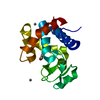
| ||||||||||||
|---|---|---|---|---|---|---|---|---|---|---|---|---|---|
| 1 |
| ||||||||||||
| Unit cell |
| ||||||||||||
| Components on special symmetry positions |
|
-Components
| #1: Protein | Mass: 23861.832 Da / Num. of mol.: 1 Source method: isolated from a genetically manipulated source Source: (gene. exp.) Thermothielavioides terrestris (fungus)Strain: ATCC 38088 / NRRL 8126 / Gene: THITE_2110902 / Production host: | ||||||
|---|---|---|---|---|---|---|---|
| #2: Chemical | ChemComp-EDO /   Mass: 62.068 Da / Num. of mol.: 1 / Source method: obtained synthetically / Formula: C2H6O2 Mass: 62.068 Da / Num. of mol.: 1 / Source method: obtained synthetically / Formula: C2H6O2 | ||||||
| #3: Chemical |   Mass: 65.409 Da / Num. of mol.: 2 / Source method: obtained synthetically / Formula: Zn Mass: 65.409 Da / Num. of mol.: 2 / Source method: obtained synthetically / Formula: Zn#4: Water | ChemComp-HOH / |  Mass: 18.015 Da / Num. of mol.: 40 / Source method: isolated from a natural source / Formula: H2O Mass: 18.015 Da / Num. of mol.: 40 / Source method: isolated from a natural source / Formula: H2OHas ligand of interest | N | Has protein modification | Y | |
-Experimental details
-Experiment
| Experiment | Method: X-RAY DIFFRACTION / Number of used crystals: 1 |
|---|
- Sample preparation
| Crystal | Density Matthews: 3.07 Å3/Da / Density % sol: 59.95 % |
|---|---|
| Crystal grow | Temperature: 293 K / Method: vapor diffusion, sitting drop Details: PACT screen condition D12 (0.01 M zinc chloride, 0.1 M Tris pH 8, 20% w/v PEG 6000) |
-Data collection
| Diffraction | Mean temperature: 100 K / Serial crystal experiment: N | ||||||||||||||||||||||||||||||
|---|---|---|---|---|---|---|---|---|---|---|---|---|---|---|---|---|---|---|---|---|---|---|---|---|---|---|---|---|---|---|---|
| Diffraction source | Source: SYNCHROTRON / Site: Diamond  / Beamline: I04-1 / Wavelength: 0.91587 Å / Beamline: I04-1 / Wavelength: 0.91587 Å | ||||||||||||||||||||||||||||||
| Detector | Type: ADSC QUANTUM 315 / Detector: CCD / Date: Apr 16, 2018 | ||||||||||||||||||||||||||||||
| Radiation | Protocol: SINGLE WAVELENGTH / Monochromatic (M) / Laue (L): M / Scattering type: x-ray | ||||||||||||||||||||||||||||||
| Radiation wavelength | Wavelength: 0.91587 Å / Relative weight: 1 | ||||||||||||||||||||||||||||||
| Reflection | Resolution: 2.36→36.64 Å / Num. obs: 10659 / % possible obs: 99.9 % / Redundancy: 12.5 % / Biso Wilson estimate: 47.4 Å2 / CC1/2: 0.999 / Rmerge(I) obs: 0.123 / Rpim(I) all: 0.051 / Rrim(I) all: 0.133 / Χ2: 1.04 / Net I/σ(I): 12.1 | ||||||||||||||||||||||||||||||
| Reflection shell | Diffraction-ID: 1
|
- Processing
| Software |
| ||||||||||||||||||||||||||||||||||||||||||||||||||||||||||||||||||||||||||||||||||||||||||||||||||||||||||||||||||||||||||||||||||||||||||||||||||||||||||||||||
|---|---|---|---|---|---|---|---|---|---|---|---|---|---|---|---|---|---|---|---|---|---|---|---|---|---|---|---|---|---|---|---|---|---|---|---|---|---|---|---|---|---|---|---|---|---|---|---|---|---|---|---|---|---|---|---|---|---|---|---|---|---|---|---|---|---|---|---|---|---|---|---|---|---|---|---|---|---|---|---|---|---|---|---|---|---|---|---|---|---|---|---|---|---|---|---|---|---|---|---|---|---|---|---|---|---|---|---|---|---|---|---|---|---|---|---|---|---|---|---|---|---|---|---|---|---|---|---|---|---|---|---|---|---|---|---|---|---|---|---|---|---|---|---|---|---|---|---|---|---|---|---|---|---|---|---|---|---|---|---|---|---|
| Refinement | Method to determine structure: MOLECULAR REPLACEMENT Starting model: 8B2E Resolution: 2.36→36.64 Å / Cor.coef. Fo:Fc: 0.969 / Cor.coef. Fo:Fc free: 0.953 / SU B: 8.739 / SU ML: 0.173 / Cross valid method: FREE R-VALUE / ESU R: 0.206 / ESU R Free: 0.174 Details: Hydrogens have been added in their riding positions
| ||||||||||||||||||||||||||||||||||||||||||||||||||||||||||||||||||||||||||||||||||||||||||||||||||||||||||||||||||||||||||||||||||||||||||||||||||||||||||||||||
| Solvent computation | Ion probe radii: 0.8 Å / Shrinkage radii: 0.8 Å / VDW probe radii: 1.2 Å / Solvent model: MASK BULK SOLVENT | ||||||||||||||||||||||||||||||||||||||||||||||||||||||||||||||||||||||||||||||||||||||||||||||||||||||||||||||||||||||||||||||||||||||||||||||||||||||||||||||||
| Displacement parameters | Biso mean: 63.097 Å2
| ||||||||||||||||||||||||||||||||||||||||||||||||||||||||||||||||||||||||||||||||||||||||||||||||||||||||||||||||||||||||||||||||||||||||||||||||||||||||||||||||
| Refinement step | Cycle: LAST / Resolution: 2.36→36.64 Å
| ||||||||||||||||||||||||||||||||||||||||||||||||||||||||||||||||||||||||||||||||||||||||||||||||||||||||||||||||||||||||||||||||||||||||||||||||||||||||||||||||
| Refine LS restraints |
| ||||||||||||||||||||||||||||||||||||||||||||||||||||||||||||||||||||||||||||||||||||||||||||||||||||||||||||||||||||||||||||||||||||||||||||||||||||||||||||||||
| LS refinement shell |
|
