 Movie
Movie Controller

[English] 日本語
 Yorodumi
- PDB-8b2d: CRYSTAL STRUCTURE OF BACTERIAL FLAVIN CONTAINING MONOOXYGENASE TH... -
+ Open data

- Basic information
| Entry | Database: PDB / ID: 8b2d | ||||||
|---|---|---|---|---|---|---|---|
| Title | CRYSTAL STRUCTURE OF BACTERIAL FLAVIN CONTAINING MONOOXYGENASE THERMORESISTANT MUTANT, IN COMPLEX WITH NADP+ | ||||||
 Components Components | Flavin-containing monooxygenase, Fmo | ||||||
 Keywords Keywords | OXIDOREDUCTASE / thermoresistant mutant / complex / flavin adenine dinucleotide / nicotinamide adenine dinucleotide phosphate | ||||||
| Function / homology |  Function and homology information Function and homology informationhypotaurine monooxygenase activity / trimethylamine monooxygenase / N,N-dimethylaniline monooxygenase activity / flavin adenine dinucleotide binding / NADP binding Similarity search - Function | ||||||
| Biological species |  Methylophaga aminisulfidivorans MP (bacteria) Methylophaga aminisulfidivorans MP (bacteria) | ||||||
| Method |  X-RAY DIFFRACTION / SYNCHROTRON / MOLECULAR REPLACEMENT / Resolution: 1.62 Å X-RAY DIFFRACTION / SYNCHROTRON / MOLECULAR REPLACEMENT / Resolution: 1.62 Å | ||||||
 Authors Authors | Cea-Rama, I. / Sanz-Aparicio, J. / Ferrer Martinez, M. / Goris, M. / Bjerga, G. | ||||||
| Funding support |  Spain, 1items Spain, 1items
| ||||||
 Citation Citation | Journal: Appl.Environ.Microbiol. / Year: 2023 Title: Increased Thermostability of an Engineered Flavin-Containing Monooxygenase to Remediate Trimethylamine in Fish Protein Hydrolysates. Authors: Goris, M. / Cea-Rama, I. / Puntervoll, P. / Ree, R. / Almendral, D. / Sanz-Aparicio, J. / Ferrer, M. / Bjerga, G.E.K. | ||||||
| History |
|
- Structure visualization
| Structure viewer | Molecule: MolmilJmol/JSmol |
|---|
- Downloads & links
-Download
| PDBx/mmCIF format | 8b2d.cif.gz | 211.4 KB | Display | PDBx/mmCIF format |
|---|---|---|---|---|
| PDB format | pdb8b2d.ent.gz | 165.7 KB | Display | PDB format |
| PDBx/mmJSON format | 8b2d.json.gz | Tree view | PDBx/mmJSON format | |
| Others |  Other downloads Other downloads |
-Validation report
| Summary document | 8b2d_validation.pdf.gz | 1.5 MB | Display | wwPDB validaton report |
|---|---|---|---|---|
| Full document | 8b2d_full_validation.pdf.gz | 1.5 MB | Display | |
| Data in XML | 8b2d_validation.xml.gz | 38.1 KB | Display | |
| Data in CIF | 8b2d_validation.cif.gz | 55.3 KB | Display | |
| Arichive directory | https://data.pdbj.org/pub/pdb/validation_reports/b2/8b2dftp://data.pdbj.org/pub/pdb/validation_reports/b2/8b2d | HTTPS FTP |
-Related structure data
| Related structure data |  2xveS S: Starting model for refinement |
|---|---|
| Similar structure data |
-Links
 PDBj
PDBj- Assembly
| Deposited unit | 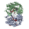
| ||||||||||||||||||
|---|---|---|---|---|---|---|---|---|---|---|---|---|---|---|---|---|---|---|---|
| 1 |
| ||||||||||||||||||
| Unit cell |
| ||||||||||||||||||
| Components on special symmetry positions |
| ||||||||||||||||||
| Noncrystallographic symmetry (NCS) | NCS domain:
NCS domain segments: Component-ID: _ / Ens-ID: 1 / Beg auth comp-ID: ALA / Beg label comp-ID: ALA / End auth comp-ID: LEU / End label comp-ID: LEU / Refine code: _ / Auth seq-ID: 2 - 444 / Label seq-ID: 2 - 444
|
-Components
-Protein , 1 types, 2 molecules AB
| #1: Protein | Mass: 54131.293 Da / Num. of mol.: 2 Source method: isolated from a genetically manipulated source Source: (gene. exp.) Methylophaga aminisulfidivorans MP (bacteria)Gene: MAMP_00532 / Production host: |
|---|
-Non-polymers , 7 types, 510 molecules 


| #2: Chemical |  Mass: 96.063 Da / Num. of mol.: 2 / Source method: obtained synthetically / Formula: SO4 Mass: 96.063 Da / Num. of mol.: 2 / Source method: obtained synthetically / Formula: SO4#3: Chemical | ChemComp-GOL /  Mass: 92.094 Da / Num. of mol.: 6 / Source method: obtained synthetically / Formula: C3H8O3 Mass: 92.094 Da / Num. of mol.: 6 / Source method: obtained synthetically / Formula: C3H8O3#4: Chemical |  Mass: 785.550 Da / Num. of mol.: 2 / Source method: obtained synthetically / Formula: C27H33N9O15P2 / Feature type: SUBJECT OF INVESTIGATION / Comment: FAD*YM Mass: 785.550 Da / Num. of mol.: 2 / Source method: obtained synthetically / Formula: C27H33N9O15P2 / Feature type: SUBJECT OF INVESTIGATION / Comment: FAD*YM#5: Chemical |  Mass: 743.405 Da / Num. of mol.: 2 / Source method: obtained synthetically / Formula: C21H28N7O17P3 / Feature type: SUBJECT OF INVESTIGATION Mass: 743.405 Da / Num. of mol.: 2 / Source method: obtained synthetically / Formula: C21H28N7O17P3 / Feature type: SUBJECT OF INVESTIGATION#6: Chemical | ChemComp-BTB / |  Mass: 209.240 Da / Num. of mol.: 1 / Source method: obtained synthetically / Formula: C8H19NO5 / Comment: pH buffer*YM Mass: 209.240 Da / Num. of mol.: 1 / Source method: obtained synthetically / Formula: C8H19NO5 / Comment: pH buffer*YM#7: Chemical |  Mass: 35.453 Da / Num. of mol.: 2 / Source method: obtained synthetically / Formula: Cl Mass: 35.453 Da / Num. of mol.: 2 / Source method: obtained synthetically / Formula: Cl#8: Water | ChemComp-HOH / | Mass: 18.015 Da / Num. of mol.: 495 / Source method: isolated from a natural source / Formula: H2O |
|---|
-Details
| Has ligand of interest | Y |
|---|
-Experimental details
-Experiment
| Experiment | Method: X-RAY DIFFRACTION / Number of used crystals: 1 |
|---|
- Sample preparation
| Crystal | Density Matthews: 2.19 Å3/Da / Density % sol: 43.85 % |
|---|---|
| Crystal grow | Temperature: 291 K / Method: vapor diffusion, sitting drop / Details: 2M (NH4)SO4, 0.1M Bis-Tris pH 6.5 |
-Data collection
| Diffraction | Mean temperature: 100 K / Serial crystal experiment: N |
|---|---|
| Diffraction source | Source: SYNCHROTRON / Site: ALBA / Beamline: XALOC / Wavelength: 0.979264 Å |
| Detector | Type: DECTRIS PILATUS3 S 6M / Detector: PIXEL / Date: May 12, 2020 / Details: KB focusing mirrors |
| Radiation | Monochromator: Si(111) channel-cut, cryocooled / Protocol: SINGLE WAVELENGTH / Monochromatic (M) / Laue (L): M / Scattering type: x-ray |
| Radiation wavelength | Wavelength: 0.979264 Å / Relative weight: 1 |
| Reflection | Resolution: 1.62→45.27 Å / Num. obs: 120046 / % possible obs: 100 % / Redundancy: 10.9 % / CC1/2: 0.999 / Rmerge(I) obs: 0.065 / Rpim(I) all: 0.021 / Net I/σ(I): 20.7 |
| Reflection shell | Resolution: 1.62→1.65 Å / Rmerge(I) obs: 0.701 / Mean I/σ(I) obs: 4.1 / Num. unique obs: 5926 / CC1/2: 0.898 / Rpim(I) all: 0.22 |
- Processing
| Software |
| ||||||||||||||||||||||||||||||||||||||||||||||||||||||||||||
|---|---|---|---|---|---|---|---|---|---|---|---|---|---|---|---|---|---|---|---|---|---|---|---|---|---|---|---|---|---|---|---|---|---|---|---|---|---|---|---|---|---|---|---|---|---|---|---|---|---|---|---|---|---|---|---|---|---|---|---|---|---|
| Refinement | Method to determine structure: MOLECULAR REPLACEMENT Starting model: 2XVE Resolution: 1.62→45.27 Å / Cor.coef. Fo:Fc: 0.971 / Cor.coef. Fo:Fc free: 0.965 / SU B: 1.505 / SU ML: 0.052 / Cross valid method: THROUGHOUT / σ(F): 0 / ESU R: 0.084 / ESU R Free: 0.079 / Stereochemistry target values: MAXIMUM LIKELIHOOD Details: HYDROGENS HAVE BEEN ADDED IN THE RIDING POSITIONS U VALUES : REFINED INDIVIDUALLY
| ||||||||||||||||||||||||||||||||||||||||||||||||||||||||||||
| Solvent computation | Ion probe radii: 0.8 Å / Shrinkage radii: 0.8 Å / VDW probe radii: 1.2 Å / Solvent model: MASK | ||||||||||||||||||||||||||||||||||||||||||||||||||||||||||||
| Displacement parameters | Biso max: 60.57 Å2 / Biso mean: 21.524 Å2 / Biso min: 12.79 Å2
| ||||||||||||||||||||||||||||||||||||||||||||||||||||||||||||
| Refinement step | Cycle: final / Resolution: 1.62→45.27 Å
| ||||||||||||||||||||||||||||||||||||||||||||||||||||||||||||
| Refine LS restraints |
| ||||||||||||||||||||||||||||||||||||||||||||||||||||||||||||
| Refine LS restraints NCS | Ens-ID: 1 / Number: 15825 / Refine-ID: X-RAY DIFFRACTION / Type: interatomic distance / Rms dev position: 0.05 Å / Weight position: 0.05
| ||||||||||||||||||||||||||||||||||||||||||||||||||||||||||||
| LS refinement shell | Resolution: 1.62→1.662 Å / Rfactor Rfree error: 0 / Total num. of bins used: 20
|
