 Movie
Movie Controller


+ Open data

- Basic information
| Entry | Database: PDB / ID: 8b1g | ||||||
|---|---|---|---|---|---|---|---|
| Title | DtpB-Nb132-AW | ||||||
 Components Components |
| ||||||
 Keywords Keywords | MEMBRANE PROTEIN / MFS / Proton coupled Oligopeptide Transporter | ||||||
| Function / homology |  Function and homology information Function and homology informationdipeptide transmembrane transport / tripeptide transmembrane transport / tripeptide transmembrane transporter activity / peptide:proton symporter activity / dipeptide transmembrane transporter activity / proton transmembrane transporter activity / proton transmembrane transport / protein transport / plasma membrane Similarity search - Function | ||||||
| Biological species |   | ||||||
| Method |  X-RAY DIFFRACTION / SYNCHROTRON / MOLECULAR REPLACEMENT / Resolution: 2.5 Å X-RAY DIFFRACTION / SYNCHROTRON / MOLECULAR REPLACEMENT / Resolution: 2.5 Å | ||||||
 Authors Authors | Killer, M. / Finocchio, G. / Lei, J. / Jungnickel, K. / Kotov, V. / Steinke, J. / Bartels, K. / Strauss, J. / Dupeux, F. / Humm, A.S. ...Killer, M. / Finocchio, G. / Lei, J. / Jungnickel, K. / Kotov, V. / Steinke, J. / Bartels, K. / Strauss, J. / Dupeux, F. / Humm, A.S. / Cornaciu, I. / Marquez, J. / Pardon, E. / Steyeart, J. / Loew, C. | ||||||
| Funding support | 1items
| ||||||
 Citation Citation | Journal: Cell Rep / Year: 2023 Title: Plasticity of the binding pocket in peptide transporters underpins promiscuous substrate recognition. Authors: Kotov, V. / Killer, M. / Jungnickel, K.E.J. / Lei, J. / Finocchio, G. / Steinke, J. / Bartels, K. / Strauss, J. / Dupeux, F. / Humm, A.S. / Cornaciu, I. / Marquez, J.A. / Pardon, E. / Steyaert, J. / Low, C. | ||||||
| History |
|
- Structure visualization
| Structure viewer | Molecule: MolmilJmol/JSmol |
|---|
- Downloads & links
-Download
| PDBx/mmCIF format | 8b1g.cif.gz | 148.8 KB | Display | PDBx/mmCIF format |
|---|---|---|---|---|
| PDB format | pdb8b1g.ent.gz | 102.8 KB | Display | PDB format |
| PDBx/mmJSON format | 8b1g.json.gz | Tree view | PDBx/mmJSON format | |
| Others |  Other downloads Other downloads |
-Validation report
| Arichive directory | https://data.pdbj.org/pub/pdb/validation_reports/b1/8b1gftp://data.pdbj.org/pub/pdb/validation_reports/b1/8b1g | HTTPS FTP |
|---|
-Related structure data
| Related structure data |  8b17C  8b18C  8b19C  8b1aC  8b1bC  8b1cC 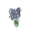 8b1dC  8b1eC 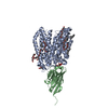 8b1fC  8b1hC 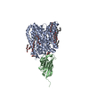 8b1iC 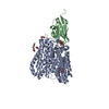 8b1jC  8b1kC  6gs4S S: Starting model for refinement C: citing same article ( |
|---|---|
| Similar structure data |
-Links
 PDBj
PDBj


- Assembly
| Deposited unit | 
| ||||||||||||
|---|---|---|---|---|---|---|---|---|---|---|---|---|---|
| 1 |
| ||||||||||||
| Unit cell |
|
-Components
-Protein / Antibody / Protein/peptide / Sugars , 4 types, 4 molecules ABC
| #1: Protein | Mass: 53607.746 Da / Num. of mol.: 1 Source method: isolated from a genetically manipulated source Source: (gene. exp.) |
|---|---|
| #2: Antibody | Mass: 13788.445 Da / Num. of mol.: 1 Source method: isolated from a genetically manipulated source Source: (gene. exp.) |
| #3: Protein/peptide | Mass: 275.303 Da / Num. of mol.: 1 / Source method: obtained synthetically / Source: (synth.) |
| #4: Sugar | ChemComp-LMT /  Type: D-saccharide / Mass: 510.615 Da / Num. of mol.: 1 / Source method: obtained synthetically / Formula: C24H46O11 / Feature type: SUBJECT OF INVESTIGATION / Comment: detergent*YM Type: D-saccharide / Mass: 510.615 Da / Num. of mol.: 1 / Source method: obtained synthetically / Formula: C24H46O11 / Feature type: SUBJECT OF INVESTIGATION / Comment: detergent*YM |
-Non-polymers , 8 types, 138 molecules 


| #5: Chemical | ChemComp-OCT /  Mass: 114.229 Da / Num. of mol.: 1 / Source method: obtained synthetically / Formula: C8H18 / Feature type: SUBJECT OF INVESTIGATION Mass: 114.229 Da / Num. of mol.: 1 / Source method: obtained synthetically / Formula: C8H18 / Feature type: SUBJECT OF INVESTIGATION | ||||||||||||
|---|---|---|---|---|---|---|---|---|---|---|---|---|---|
| #6: Chemical | ChemComp-PG4 /  Mass: 194.226 Da / Num. of mol.: 5 / Source method: obtained synthetically / Formula: C8H18O5 / Feature type: SUBJECT OF INVESTIGATION / Comment: precipitant*YM Mass: 194.226 Da / Num. of mol.: 5 / Source method: obtained synthetically / Formula: C8H18O5 / Feature type: SUBJECT OF INVESTIGATION / Comment: precipitant*YM#7: Chemical |  Mass: 238.278 Da / Num. of mol.: 2 / Source method: obtained synthetically / Formula: C10H22O6 / Feature type: SUBJECT OF INVESTIGATION / Comment: precipitant*YM Mass: 238.278 Da / Num. of mol.: 2 / Source method: obtained synthetically / Formula: C10H22O6 / Feature type: SUBJECT OF INVESTIGATION / Comment: precipitant*YM#8: Chemical | ChemComp-D12 / |  Mass: 170.335 Da / Num. of mol.: 1 / Source method: obtained synthetically / Formula: C12H26 / Feature type: SUBJECT OF INVESTIGATION Mass: 170.335 Da / Num. of mol.: 1 / Source method: obtained synthetically / Formula: C12H26 / Feature type: SUBJECT OF INVESTIGATION#9: Chemical |  Mass: 92.094 Da / Num. of mol.: 2 / Source method: obtained synthetically / Formula: C3H8O3 / Feature type: SUBJECT OF INVESTIGATION Mass: 92.094 Da / Num. of mol.: 2 / Source method: obtained synthetically / Formula: C3H8O3 / Feature type: SUBJECT OF INVESTIGATION#10: Chemical | ChemComp-D10 / |  Mass: 142.282 Da / Num. of mol.: 1 / Source method: obtained synthetically / Formula: C10H22 / Feature type: SUBJECT OF INVESTIGATION Mass: 142.282 Da / Num. of mol.: 1 / Source method: obtained synthetically / Formula: C10H22 / Feature type: SUBJECT OF INVESTIGATION#11: Chemical |  Mass: 86.175 Da / Num. of mol.: 2 / Source method: obtained synthetically / Formula: C6H14 / Feature type: SUBJECT OF INVESTIGATION Mass: 86.175 Da / Num. of mol.: 2 / Source method: obtained synthetically / Formula: C6H14 / Feature type: SUBJECT OF INVESTIGATION#12: Water | ChemComp-HOH / | Mass: 18.015 Da / Num. of mol.: 124 / Source method: isolated from a natural source / Formula: H2O |
-Details
| Has ligand of interest | Y |
|---|---|
| Has protein modification | Y |
-Experimental details
-Experiment
| Experiment | Method: X-RAY DIFFRACTION / Number of used crystals: 1 |
|---|
- Sample preparation
| Crystal | Density Matthews: 4.29 Å3/Da / Density % sol: 71.3 % |
|---|---|
| Crystal grow | Temperature: 292 K / Method: vapor diffusion Details: 100 mM HEPES pH 7.0, 30-40 % PEG 400 100-300 mM NaCl, 200 mM Li2SO4 and 10 mM AW |
-Data collection
| Diffraction | Mean temperature: 100 K / Serial crystal experiment: N |
|---|---|
| Diffraction source | Source: SYNCHROTRON / Site: PETRA III, EMBL c/o DESY  / Beamline: P13 (MX1) / Wavelength: 0.98 Å / Beamline: P13 (MX1) / Wavelength: 0.98 Å |
| Detector | Type: DECTRIS EIGER X 16M / Detector: PIXEL / Date: May 19, 2020 |
| Radiation | Protocol: SINGLE WAVELENGTH / Monochromatic (M) / Laue (L): M / Scattering type: x-ray |
| Radiation wavelength | Wavelength: 0.98 Å / Relative weight: 1 |
| Reflection | Resolution: 2.5→69.97 Å / Num. obs: 77038 / % possible obs: 99.15 % / Redundancy: 2 % / Biso Wilson estimate: 69.68 Å2 / CC1/2: 1 / Net I/σ(I): 12.36 |
| Reflection shell | Resolution: 2.5→2.589 Å / Num. unique obs: 4000 / CC1/2: 0.432 |
- Processing
| Software | Name: PHENIX / Version: 1.19.2_4158 / Classification: refinement | ||||||||||||||||||||||||||||||||||||||||||||||||||||||||||||||||||||||||||||||||||||||||||||||||||||||||||||||||||||||||||||||||||||||||||||||||||||||||||||||||||||||||||||||||||||||||||||||||||||
|---|---|---|---|---|---|---|---|---|---|---|---|---|---|---|---|---|---|---|---|---|---|---|---|---|---|---|---|---|---|---|---|---|---|---|---|---|---|---|---|---|---|---|---|---|---|---|---|---|---|---|---|---|---|---|---|---|---|---|---|---|---|---|---|---|---|---|---|---|---|---|---|---|---|---|---|---|---|---|---|---|---|---|---|---|---|---|---|---|---|---|---|---|---|---|---|---|---|---|---|---|---|---|---|---|---|---|---|---|---|---|---|---|---|---|---|---|---|---|---|---|---|---|---|---|---|---|---|---|---|---|---|---|---|---|---|---|---|---|---|---|---|---|---|---|---|---|---|---|---|---|---|---|---|---|---|---|---|---|---|---|---|---|---|---|---|---|---|---|---|---|---|---|---|---|---|---|---|---|---|---|---|---|---|---|---|---|---|---|---|---|---|---|---|---|---|---|---|
| Refinement | Method to determine structure: MOLECULAR REPLACEMENT Starting model: 6gs4 Resolution: 2.5→69.97 Å / SU ML: 0.3661 / Cross valid method: FREE R-VALUE / σ(F): 1.33 / Phase error: 27.1074 Stereochemistry target values: GeoStd + Monomer Library + CDL v1.2
| ||||||||||||||||||||||||||||||||||||||||||||||||||||||||||||||||||||||||||||||||||||||||||||||||||||||||||||||||||||||||||||||||||||||||||||||||||||||||||||||||||||||||||||||||||||||||||||||||||||
| Solvent computation | Shrinkage radii: 0.9 Å / VDW probe radii: 1.11 Å / Solvent model: FLAT BULK SOLVENT MODEL | ||||||||||||||||||||||||||||||||||||||||||||||||||||||||||||||||||||||||||||||||||||||||||||||||||||||||||||||||||||||||||||||||||||||||||||||||||||||||||||||||||||||||||||||||||||||||||||||||||||
| Displacement parameters | Biso mean: 77.25 Å2 | ||||||||||||||||||||||||||||||||||||||||||||||||||||||||||||||||||||||||||||||||||||||||||||||||||||||||||||||||||||||||||||||||||||||||||||||||||||||||||||||||||||||||||||||||||||||||||||||||||||
| Refinement step | Cycle: LAST / Resolution: 2.5→69.97 Å
| ||||||||||||||||||||||||||||||||||||||||||||||||||||||||||||||||||||||||||||||||||||||||||||||||||||||||||||||||||||||||||||||||||||||||||||||||||||||||||||||||||||||||||||||||||||||||||||||||||||
| Refine LS restraints |
| ||||||||||||||||||||||||||||||||||||||||||||||||||||||||||||||||||||||||||||||||||||||||||||||||||||||||||||||||||||||||||||||||||||||||||||||||||||||||||||||||||||||||||||||||||||||||||||||||||||
| LS refinement shell |
|
