 Movie
Movie Controller


+ Open data

- Basic information
| Entry | Database: PDB / ID: 8b0u | ||||||
|---|---|---|---|---|---|---|---|
| Title | Structure of the CalpL/T10 complex | ||||||
 Components Components |
| ||||||
 Keywords Keywords | HYDROLASE / Protease / CRISPR type III / cOA / cA4 / peptide binding protein | ||||||
| Function / homology | SMODS-associated and fused to various effectors / SMODS-associated and fused to various effectors sensor domain / Ribosomal protein S5 domain 2-type fold, subgroup / Ribosomal protein S5 domain 2-type fold / membrane / DUF4388 domain-containing protein / SMODS-associated and fused to various effectors domain-containing protein Function and homology information Function and homology information | ||||||
| Biological species |  Sulfurihydrogenibium (bacteria) Sulfurihydrogenibium (bacteria) | ||||||
| Method |  X-RAY DIFFRACTION / SYNCHROTRON / MOLECULAR REPLACEMENT / Resolution: 3.29 Å X-RAY DIFFRACTION / SYNCHROTRON / MOLECULAR REPLACEMENT / Resolution: 3.29 Å | ||||||
 Authors Authors | Schneberger, N. / Hagelueken, G. | ||||||
| Funding support |  Germany, 1items Germany, 1items
| ||||||
 Citation Citation | Journal: Nature / Year: 2023 Title: Antiviral signalling by a cyclic nucleotide activated CRISPR protease. Authors: Rouillon, C. / Schneberger, N. / Chi, H. / Blumenstock, K. / Da Vela, S. / Ackermann, K. / Moecking, J. / Peter, M.F. / Boenigk, W. / Seifert, R. / Bode, B.E. / Schmid-Burgk, J.L. / Svergun, ...Authors: Rouillon, C. / Schneberger, N. / Chi, H. / Blumenstock, K. / Da Vela, S. / Ackermann, K. / Moecking, J. / Peter, M.F. / Boenigk, W. / Seifert, R. / Bode, B.E. / Schmid-Burgk, J.L. / Svergun, D. / Geyer, M. / White, M.F. / Hagelueken, G. | ||||||
| History |
|
- Structure visualization
| Structure viewer | Molecule: MolmilJmol/JSmol |
|---|
- Downloads & links
-Download
| PDBx/mmCIF format | 8b0u.cif.gz | 241.2 KB | Display | PDBx/mmCIF format |
|---|---|---|---|---|
| PDB format | pdb8b0u.ent.gz | 193 KB | Display | PDB format |
| PDBx/mmJSON format | 8b0u.json.gz | Tree view | PDBx/mmJSON format | |
| Others |  Other downloads Other downloads |
-Validation report
| Arichive directory | https://data.pdbj.org/pub/pdb/validation_reports/b0/8b0uftp://data.pdbj.org/pub/pdb/validation_reports/b0/8b0u | HTTPS FTP |
|---|
-Related structure data
| Related structure data | 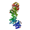 7qdaSC 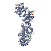 8b0rC S: Starting model for refinement C: citing same article ( |
|---|---|
| Similar structure data | |
| Other databases |
|
-Links
 PDBj
PDBj- Assembly
| Deposited unit | 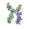
| ||||||||
|---|---|---|---|---|---|---|---|---|---|
| 1 | 
| ||||||||
| 2 | 
| ||||||||
| Unit cell |
|
-Components
| #1: Protein | Mass: 57813.680 Da / Num. of mol.: 2 Source method: isolated from a genetically manipulated source Details: CalpL protease / Source: (gene. exp.) Sulfurihydrogenibium (bacteria) / Strain: YO3AOP1 / Gene: SYO3AOP1_0656 / Production host: #2: Protein | Mass: 8755.967 Da / Num. of mol.: 2 Source method: isolated from a genetically manipulated source Details: This is the C-terminal fragment of CalpT that remains after cleavage by CalpL Source: (gene. exp.) Sulfurihydrogenibium (bacteria) / Strain: YO3AOP1 / Gene: SYO3AOP1_0655 / Production host: #3: Chemical | ChemComp-SO4 /   Mass: 96.063 Da / Num. of mol.: 7 / Source method: obtained synthetically / Formula: SO4 Mass: 96.063 Da / Num. of mol.: 7 / Source method: obtained synthetically / Formula: SO4#4: Chemical |   Mass: 92.094 Da / Num. of mol.: 2 / Source method: obtained synthetically / Formula: C3H8O3 Mass: 92.094 Da / Num. of mol.: 2 / Source method: obtained synthetically / Formula: C3H8O3#5: Water | ChemComp-HOH / |  Mass: 18.015 Da / Num. of mol.: 2 / Source method: isolated from a natural source / Formula: H2O Mass: 18.015 Da / Num. of mol.: 2 / Source method: isolated from a natural source / Formula: H2OHas ligand of interest | N | |
|---|
-Experimental details
-Experiment
| Experiment | Method: X-RAY DIFFRACTION / Number of used crystals: 1 |
|---|
- Sample preparation
| Crystal | Density Matthews: 4.24 Å3/Da / Density % sol: 71.02 % |
|---|---|
| Crystal grow | Temperature: 293 K / Method: vapor diffusion, sitting drop Details: 2 M ammonium sulfate, 0.1 M ammonium cacodylate, 0.2 M NaCl at pH 6.5 |
-Data collection
| Diffraction | Mean temperature: 100 K / Serial crystal experiment: N |
|---|---|
| Diffraction source | Source: SYNCHROTRON / Site: PETRA III, EMBL c/o DESY / Beamline: P13 (MX1) / Wavelength: 0.9762 Å |
| Detector | Type: DECTRIS EIGER X 16M / Detector: PIXEL / Date: Aug 14, 2021 |
| Radiation | Protocol: SINGLE WAVELENGTH / Monochromatic (M) / Laue (L): M / Scattering type: x-ray |
| Radiation wavelength | Wavelength: 0.9762 Å / Relative weight: 1 |
| Reflection | Resolution: 3.29→49.31 Å / Num. obs: 33777 / % possible obs: 99.12 % / Redundancy: 6.8 % / CC1/2: 0.997 / Net I/σ(I): 9.92 |
| Reflection shell | Resolution: 3.29→3.408 Å / Redundancy: 6.9 % / Num. unique obs: 3321 / CC1/2: 0.467 / % possible all: 99.34 |
- Processing
| Software | Name: PHASER / Classification: phasing | |||||||||||||||||||||||||||||||||||||||||||||||||||||||||||||||||||||||||||||||||||||||||||||||||||||||||
|---|---|---|---|---|---|---|---|---|---|---|---|---|---|---|---|---|---|---|---|---|---|---|---|---|---|---|---|---|---|---|---|---|---|---|---|---|---|---|---|---|---|---|---|---|---|---|---|---|---|---|---|---|---|---|---|---|---|---|---|---|---|---|---|---|---|---|---|---|---|---|---|---|---|---|---|---|---|---|---|---|---|---|---|---|---|---|---|---|---|---|---|---|---|---|---|---|---|---|---|---|---|---|---|---|---|---|
| Refinement | Method to determine structure: MOLECULAR REPLACEMENT Starting model: 7QDA Resolution: 3.29→49.31 Å / SU ML: 0.45 / Cross valid method: FREE R-VALUE / σ(F): 1.34 / Phase error: 25.69 / Stereochemistry target values: ML
| |||||||||||||||||||||||||||||||||||||||||||||||||||||||||||||||||||||||||||||||||||||||||||||||||||||||||
| Solvent computation | Shrinkage radii: 0.9 Å / VDW probe radii: 1.11 Å / Solvent model: FLAT BULK SOLVENT MODEL | |||||||||||||||||||||||||||||||||||||||||||||||||||||||||||||||||||||||||||||||||||||||||||||||||||||||||
| Refinement step | Cycle: LAST / Resolution: 3.29→49.31 Å
| |||||||||||||||||||||||||||||||||||||||||||||||||||||||||||||||||||||||||||||||||||||||||||||||||||||||||
| Refine LS restraints |
| |||||||||||||||||||||||||||||||||||||||||||||||||||||||||||||||||||||||||||||||||||||||||||||||||||||||||
| LS refinement shell |
|
