 Movie
Movie Controller


+ Open data

- Basic information
| Entry | Database: PDB / ID: 8az0 | |||||||||
|---|---|---|---|---|---|---|---|---|---|---|
| Title | IAPP S20G growth-phase fibril polymorph 2PF-L | |||||||||
 Components Components | Islet amyloid polypeptide | |||||||||
 Keywords Keywords | PROTEIN FIBRIL / Amyloid / fibril / helical / cross-beta / Amylin / polymorph / islet / Diabetes | |||||||||
| Function / homology |  Function and homology information Function and homology informationamylin receptor 3 signaling pathway / amylin receptor 2 signaling pathway / amylin receptor 1 signaling pathway / amylin receptor signaling pathway / Calcitonin-like ligand receptors / negative regulation of amyloid fibril formation / negative regulation of bone resorption / negative regulation of osteoclast differentiation / eating behavior / Regulation of gene expression in beta cells ...amylin receptor 3 signaling pathway / amylin receptor 2 signaling pathway / amylin receptor 1 signaling pathway / amylin receptor signaling pathway / Calcitonin-like ligand receptors / negative regulation of amyloid fibril formation / negative regulation of bone resorption / negative regulation of osteoclast differentiation / eating behavior / Regulation of gene expression in beta cells / positive regulation of cAMP/PKA signal transduction / bone resorption / negative regulation of protein-containing complex assembly / positive regulation of calcium-mediated signaling / osteoclast differentiation / sensory perception of pain / hormone activity / cell-cell signaling / amyloid-beta binding / G alpha (s) signalling events / positive regulation of MAPK cascade / positive regulation of apoptotic process / Amyloid fiber formation / receptor ligand activity / signaling receptor binding / neuronal cell body / apoptotic process / lipid binding / signal transduction / : / extracellular region / identical protein binding Similarity search - Function | |||||||||
| Biological species |  Homo sapiens (human) Homo sapiens (human) | |||||||||
| Method | ELECTRON MICROSCOPY / helical reconstruction / cryo EM / Resolution: 3.4 Å | |||||||||
 Authors Authors | Wilkinson, M. / Xu, Y. / Gallardo, R. / Radford, S.E. / Ranson, N.A. | |||||||||
| Funding support |  United Kingdom, 2items United Kingdom, 2items
| |||||||||
 Citation Citation | Journal: Cell / Year: 2023 Title: Structural evolution of fibril polymorphs during amyloid assembly. Authors: Martin Wilkinson / Yong Xu / Dev Thacker / Alexander I P Taylor / Declan G Fisher / Rodrigo U Gallardo / Sheena E Radford / Neil A Ranson / Abstract: Cryoelectron microscopy (cryo-EM) has provided unprecedented insights into amyloid fibril structures, including those associated with disease. However, these structures represent the endpoints of ...Cryoelectron microscopy (cryo-EM) has provided unprecedented insights into amyloid fibril structures, including those associated with disease. However, these structures represent the endpoints of long assembly processes, and their relationship to fibrils formed early in assembly is unknown. Consequently, whether different fibril architectures, with potentially different pathological properties, form during assembly remains unknown. Here, we used cryo-EM to determine structures of amyloid fibrils at different times during in vitro fibrillation of a disease-related variant of human islet amyloid polypeptide (IAPP-S20G). Strikingly, the fibrils formed in the lag, growth, and plateau phases have different structures, with new forms appearing and others disappearing as fibrillation proceeds. A time course with wild-type hIAPP also shows fibrils changing with time, suggesting that this is a general property of IAPP amyloid assembly. The observation of transiently populated fibril structures has implications for understanding amyloid assembly mechanisms with potential new insights into amyloid progression in disease. | |||||||||
| History |
|
- Structure visualization
| Structure viewer | Molecule: MolmilJmol/JSmol |
|---|
- Downloads & links
-Download
| PDBx/mmCIF format | 8az0.cif.gz | 57.7 KB | Display | PDBx/mmCIF format |
|---|---|---|---|---|
| PDB format | pdb8az0.ent.gz | 42.7 KB | Display | PDB format |
| PDBx/mmJSON format | 8az0.json.gz | Tree view | PDBx/mmJSON format | |
| Others |  Other downloads Other downloads |
-Validation report
| Arichive directory | https://data.pdbj.org/pub/pdb/validation_reports/az/8az0ftp://data.pdbj.org/pub/pdb/validation_reports/az/8az0 | HTTPS FTP |
|---|
-Related structure data
| Related structure data |  15728MC  8awtC 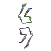 8az1C  8az2C 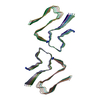 8az3C  8az4C 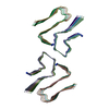 8az5C  8az6C  8az7C C: citing same article ( M: map data used to model this data |
|---|---|
| Similar structure data |


-Links
 PDBj
PDBj


- Assembly
| Deposited unit | 
|
|---|---|
| 1 |
|
-Components
| #1: Protein/peptide | Mass: 3877.286 Da / Num. of mol.: 10 / Mutation: S20G / Source method: obtained synthetically / Details: Synthesised with C-terminal amidation / Source: (synth.) Homo sapiens (human) / References: UniProt: P10997Has ligand of interest | N | Has protein modification | Y | |
|---|
-Experimental details
-Experiment
| Experiment | Method: ELECTRON MICROSCOPY |
|---|---|
| EM experiment | Aggregation state: FILAMENT / 3D reconstruction method: helical reconstruction |
- Sample preparation
| Component | Name: IAPP S20G growth-phase fibril polymorph 2PF-L / Type: COMPLEX Details: In vitro fibril growth for 6 weeks at room temperature Entity ID: all / Source: RECOMBINANT |
|---|---|
| Molecular weight | Experimental value: NO |
| Source (natural) | Organism: Homo sapiens (human) |
| Source (recombinant) | Organism: synthetic construct (others) |
| Buffer solution | pH: 6.8 |
| Buffer component | Conc.: 20 mM / Name: ammonium acetate / Formula: NH3CH3CO2 |
| Specimen | Conc.: 0.12 mg/ml / Embedding applied: NO / Shadowing applied: NO / Staining applied: NO / Vitrification applied: YES Details: Fibrillation conditions: 30 uM monomeric IAPP-S20G, quiescent at room temp for 6 weeks |
| Specimen support | Grid material: COPPER / Grid mesh size: 300 divisions/in. / Grid type: EMS Lacey Carbon |
| Vitrification | Instrument: FEI VITROBOT MARK IV / Cryogen name: ETHANE / Humidity: 90 % / Chamber temperature: 277 K / Details: 6s blot |
- Electron microscopy imaging
| Experimental equipment |  Model: Titan Krios / Image courtesy: FEI Company |
|---|---|
| Microscopy | Model: FEI TITAN KRIOS |
| Electron gun | Electron source:  FIELD EMISSION GUN / Accelerating voltage: 300 kV / Illumination mode: FLOOD BEAM FIELD EMISSION GUN / Accelerating voltage: 300 kV / Illumination mode: FLOOD BEAM |
| Electron lens | Mode: BRIGHT FIELD / Nominal magnification: 96000 X / Nominal defocus max: 2500 nm / Nominal defocus min: 1300 nm / Cs: 2.7 mm / C2 aperture diameter: 50 µm / Alignment procedure: COMA FREE |
| Specimen holder | Specimen holder model: FEI TITAN KRIOS AUTOGRID HOLDER |
| Image recording | Average exposure time: 5 sec. / Electron dose: 39 e/Å2 / Film or detector model: FEI FALCON IV (4k x 4k) / Num. of grids imaged: 1 / Num. of real images: 2350 Details: 1204 raw EER frames were collected per image and combined into 30 fractions for processing |
- Processing
| EM software |
| ||||||||||||||||||||||||||||||||||||
|---|---|---|---|---|---|---|---|---|---|---|---|---|---|---|---|---|---|---|---|---|---|---|---|---|---|---|---|---|---|---|---|---|---|---|---|---|---|
| CTF correction | Type: PHASE FLIPPING AND AMPLITUDE CORRECTION | ||||||||||||||||||||||||||||||||||||
| Helical symmerty | Angular rotation/subunit: 178.77 ° / Axial rise/subunit: 2.39 Å / Axial symmetry: C1 | ||||||||||||||||||||||||||||||||||||
| Particle selection | Num. of particles selected: 842792 Details: Manually picked a subset of images to train a model for automatic fibril segment picking in crYOLO | ||||||||||||||||||||||||||||||||||||
| 3D reconstruction | Resolution: 3.4 Å / Resolution method: FSC 0.143 CUT-OFF / Num. of particles: 12934 / Symmetry type: HELICAL | ||||||||||||||||||||||||||||||||||||
| Atomic model building | B value: 68 / Protocol: AB INITIO MODEL / Space: REAL / Target criteria: Correlation coefficient |
