 Movie
Movie Controller

[English] 日本語
 Yorodumi
- PDB-8ayg: Crystal structure of an intramolecular i-motif at the insulin-lin... -
+ Open data

- Basic information
| Entry | Database: PDB / ID: 8ayg | ||||||
|---|---|---|---|---|---|---|---|
| Title | Crystal structure of an intramolecular i-motif at the insulin-linked polymorphic region (ILPR) | ||||||
 Components Components | Insulin-linked polymorphic region, ILPR DNA (31-MER) | ||||||
 Keywords Keywords | DNA / i-motif / promoter / insulin-linked polymorphic region | ||||||
| Function / homology | DNA / DNA (> 10) Function and homology information Function and homology information | ||||||
| Biological species |  Homo sapiens (human) Homo sapiens (human) | ||||||
| Method |  X-RAY DIFFRACTION / SYNCHROTRON / MAD / Resolution: 2.25 Å X-RAY DIFFRACTION / SYNCHROTRON / MAD / Resolution: 2.25 Å | ||||||
 Authors Authors | Parkinson, G.N. / Alexandrou, E. / Waller, Z.A.E. / El-Omari, K. | ||||||
| Funding support |  United Kingdom, 1items United Kingdom, 1items
| ||||||
 Citation Citation | Journal: Nat Commun / Year: 2024 Title: Structural insights into i-motif DNA structures in sequences from the insulin-linked polymorphic region. Authors: Guneri, D. / Alexandrou, E. / El Omari, K. / Dvorakova, Z. / Chikhale, R.V. / Pike, D.T.S. / Waudby, C.A. / Morris, C.J. / Haider, S. / Parkinson, G.N. / Waller, Z.A.E. | ||||||
| History |
|
- Structure visualization
| Structure viewer | Molecule: MolmilJmol/JSmol |
|---|
- Downloads & links
-Download
| PDBx/mmCIF format | 8ayg.cif.gz | 48.7 KB | Display | PDBx/mmCIF format |
|---|---|---|---|---|
| PDB format | pdb8ayg.ent.gz | 28.6 KB | Display | PDB format |
| PDBx/mmJSON format | 8ayg.json.gz | Tree view | PDBx/mmJSON format | |
| Others |  Other downloads Other downloads |
-Validation report
| Arichive directory | https://data.pdbj.org/pub/pdb/validation_reports/ay/8aygftp://data.pdbj.org/pub/pdb/validation_reports/ay/8ayg | HTTPS FTP |
|---|
-Related structure data
| Similar structure data |
|---|
-Links
 PDBj
PDBj


- Assembly
| Deposited unit | 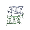
| ||||||||||
|---|---|---|---|---|---|---|---|---|---|---|---|
| 1 | 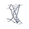
| ||||||||||
| 2 | 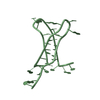
| ||||||||||
| Unit cell |
|
-Components
| #1: DNA chain | Mass: 9177.918 Da / Num. of mol.: 2 / Source method: obtained synthetically / Source: (synth.) Homo sapiens (human)#2: Water | ChemComp-HOH / |  Mass: 18.015 Da / Num. of mol.: 22 / Source method: isolated from a natural source / Formula: H2O Mass: 18.015 Da / Num. of mol.: 22 / Source method: isolated from a natural source / Formula: H2O |
|---|
-Experimental details
-Experiment
| Experiment | Method: X-RAY DIFFRACTION / Number of used crystals: 1 |
|---|
- Sample preparation
| Crystal | Density Matthews: 2.32 Å3/Da / Density % sol: 47.06 % / Description: pseudo-hexagonal |
|---|---|
| Crystal grow | Temperature: 283.15 K / Method: vapor diffusion, hanging drop / pH: 5.5 Details: 2-Methyl-2,4-pentanediol (MPD), sodium chloride, sodium cacodylate, spermine (tetrachloride) PH range: 5.5 - 6.4 / Temp details: crystals can also be formed at 277.15K |
-Data collection
| Diffraction | Mean temperature: 75 K / Ambient temp details: in vacuum (I23) / Serial crystal experiment: N | ||||||||||||||||||||||||||||||
|---|---|---|---|---|---|---|---|---|---|---|---|---|---|---|---|---|---|---|---|---|---|---|---|---|---|---|---|---|---|---|---|
| Diffraction source | Source: SYNCHROTRON / Site: Diamond / Beamline: I23 / Wavelength: 2.4797 Å | ||||||||||||||||||||||||||||||
| Detector | Type: DECTRIS PILATUS 2M / Detector: PIXEL / Date: Dec 2, 2021 | ||||||||||||||||||||||||||||||
| Radiation | Monochromator: M / Protocol: SINGLE WAVELENGTH / Monochromatic (M) / Laue (L): M / Scattering type: x-ray | ||||||||||||||||||||||||||||||
| Radiation wavelength | Wavelength: 2.4797 Å / Relative weight: 1 | ||||||||||||||||||||||||||||||
| Reflection | Resolution: 2.25→47.42 Å / Num. obs: 8133 / % possible obs: 95.2 % / Redundancy: 44.5 % / Biso Wilson estimate: 76.91 Å2 / CC1/2: 0.991 / Rmerge(I) obs: 0.167 / Rpim(I) all: 0.024 / Rrim(I) all: 0.169 / Net I/σ(I): 30.4 / Num. measured all: 362297 / Scaling rejects: 17192 | ||||||||||||||||||||||||||||||
| Reflection shell | Diffraction-ID: 1
|
-Phasing
| Phasing | Method: MAD |
|---|
- Processing
| Software |
| ||||||||||||||||||||||||||||
|---|---|---|---|---|---|---|---|---|---|---|---|---|---|---|---|---|---|---|---|---|---|---|---|---|---|---|---|---|---|
| Refinement | Method to determine structure: MAD / Resolution: 2.25→41.46 Å / SU ML: 0.5215 / Cross valid method: FREE R-VALUE / σ(F): 1.35 / Phase error: 45.592 Stereochemistry target values: GeoStd + Monomer Library + CDL v1.2
| ||||||||||||||||||||||||||||
| Solvent computation | Shrinkage radii: 0.9 Å / VDW probe radii: 1.1 Å / Solvent model: FLAT BULK SOLVENT MODEL | ||||||||||||||||||||||||||||
| Displacement parameters | Biso mean: 80.06 Å2 | ||||||||||||||||||||||||||||
| Refinement step | Cycle: LAST / Resolution: 2.25→41.46 Å
| ||||||||||||||||||||||||||||
| Refine LS restraints |
| ||||||||||||||||||||||||||||
| LS refinement shell |
|
