 Movie
Movie Controller

[English] 日本語
 Yorodumi
- PDB-8axo: Crystal structure of the C-terminal domain of Trypanosoma brucei ... -
+ Open data

- Basic information
| Entry | Database: PDB / ID: 8axo | ||||||
|---|---|---|---|---|---|---|---|
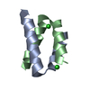
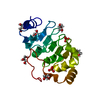
| Title | Crystal structure of the C-terminal domain of Trypanosoma brucei CFAP410 | ||||||
 Components Components | TbCFAP410-CTD | ||||||
 Keywords Keywords | STRUCTURAL PROTEIN / Cilia / Flagella / Coiled-coil | ||||||
| Function / homology | : / CFAP410 C-terminal domain / cell tip / Leucine-rich repeat / Leucine-rich repeat profile. / Leucine-rich repeat / Leucine-rich repeat domain superfamily / cytoplasm / CFAP410 C-terminal domain-containing protein Function and homology information Function and homology information | ||||||
| Biological species |  | ||||||
| Method |  X-RAY DIFFRACTION / SYNCHROTRON / AB INITIO PHASING / Resolution: 1.29 Å X-RAY DIFFRACTION / SYNCHROTRON / AB INITIO PHASING / Resolution: 1.29 Å | ||||||
 Authors Authors | Dong, G. / Stadler, A. | ||||||
| Funding support |  Austria, 1items Austria, 1items
| ||||||
 Citation Citation | Journal: Open Biology / Year: 2024 Title: The C-terminus of CFAP410 forms a tetrameric helical bundle that is essential for its localization to the basal body. Authors: Stadler, A. / De Liz, L.V. / Gabriel, H.B. / Alonso-Gil, S. / Crickley, R. / Korbula, K. / Zagrovic, B. / Vaughan, S. / Sunter, J.D. / Dong, G. | ||||||
| History |
|
- Structure visualization
| Structure viewer | Molecule: MolmilJmol/JSmol |
|---|
- Downloads & links
-Download
| PDBx/mmCIF format | 8axo.cif.gz | 59 KB | Display | PDBx/mmCIF format |
|---|---|---|---|---|
| PDB format | pdb8axo.ent.gz | 37 KB | Display | PDB format |
| PDBx/mmJSON format | 8axo.json.gz | Tree view | PDBx/mmJSON format | |
| Others |  Other downloads Other downloads |
-Validation report
| Arichive directory | https://data.pdbj.org/pub/pdb/validation_reports/ax/8axoftp://data.pdbj.org/pub/pdb/validation_reports/ax/8axo | HTTPS FTP |
|---|
-Related structure data

-Links
 PDBj
PDBj


- Assembly
| Deposited unit | 
| |||||||||||||||||||||||||||||||||||||||||||||||||||||||||||||||||||||||||||||||
|---|---|---|---|---|---|---|---|---|---|---|---|---|---|---|---|---|---|---|---|---|---|---|---|---|---|---|---|---|---|---|---|---|---|---|---|---|---|---|---|---|---|---|---|---|---|---|---|---|---|---|---|---|---|---|---|---|---|---|---|---|---|---|---|---|---|---|---|---|---|---|---|---|---|---|---|---|---|---|---|---|
| 1 | 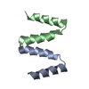
| |||||||||||||||||||||||||||||||||||||||||||||||||||||||||||||||||||||||||||||||
| Unit cell |
| |||||||||||||||||||||||||||||||||||||||||||||||||||||||||||||||||||||||||||||||
| Noncrystallographic symmetry (NCS) | NCS domain:
NCS domain segments: Ens-ID: ens_1
NCS oper: (Code: givenMatrix: (0.414888869432, -0.817997484866, -0.398431099156), (-0.816003137042, -0.528227217356, 0.234765600509), (-0.402499821565, 0.227719392229, -0.886644106755)Vector: -20. ...NCS oper: (Code: given Matrix: (0.414888869432, -0.817997484866, -0.398431099156), Vector: |
-Components
| #1: Protein/peptide | Mass: 3913.451 Da / Num. of mol.: 2 Source method: isolated from a genetically manipulated source Source: (gene. exp.) Strain: 927/4 GUTat10.1 / Gene: Tb09.211.3240 / Production host:  #2: Water | ChemComp-HOH / |  Mass: 18.015 Da / Num. of mol.: 33 / Source method: isolated from a natural source / Formula: H2O Mass: 18.015 Da / Num. of mol.: 33 / Source method: isolated from a natural source / Formula: H2O |
|---|
-Experimental details
-Experiment
| Experiment | Method: X-RAY DIFFRACTION / Number of used crystals: 1 |
|---|
- Sample preparation
| Crystal | Density Matthews: 1.86 Å3/Da / Density % sol: 33.85 % |
|---|---|
| Crystal grow | Temperature: 277 K / Method: evaporation / Details: 2.0 M ammonium sulfate, 5% (v/v) iso-propanol / PH range: 7.0-7.5 |
-Data collection
| Diffraction | Mean temperature: 100 K / Serial crystal experiment: N |
|---|---|
| Diffraction source | Source: SYNCHROTRON / Site: ESRF  / Beamline: ID23-1 / Wavelength: 0.97242 Å / Beamline: ID23-1 / Wavelength: 0.97242 Å |
| Detector | Type: DECTRIS PILATUS 6M / Detector: PIXEL / Date: Nov 6, 2020 |
| Radiation | Protocol: SINGLE WAVELENGTH / Monochromatic (M) / Laue (L): M / Scattering type: x-ray |
| Radiation wavelength | Wavelength: 0.97242 Å / Relative weight: 1 |
| Reflection | Resolution: 1.29→19.1 Å / Num. obs: 15025 / % possible obs: 99.65 % / Redundancy: 9.4 % / Biso Wilson estimate: 22.58 Å2 / CC1/2: 0.991 / CC star: 0.998 / Rmerge(I) obs: 0.132 / Rpim(I) all: 0.046 / Rrim(I) all: 0.14 / Net I/σ(I): 8.16 |
| Reflection shell | Resolution: 1.29→1.34 Å / Num. unique obs: 1430 / CC1/2: 0.43 / CC star: 0.775 |
- Processing
| Software |
| ||||||||||||||||||||||||||||||||||||||||||||||||||||||||||||||||||||||||||||||||||||
|---|---|---|---|---|---|---|---|---|---|---|---|---|---|---|---|---|---|---|---|---|---|---|---|---|---|---|---|---|---|---|---|---|---|---|---|---|---|---|---|---|---|---|---|---|---|---|---|---|---|---|---|---|---|---|---|---|---|---|---|---|---|---|---|---|---|---|---|---|---|---|---|---|---|---|---|---|---|---|---|---|---|---|---|---|---|
| Refinement | Method to determine structure: AB INITIO PHASING / Resolution: 1.29→19.1 Å / SU ML: 0.1764 / Cross valid method: FREE R-VALUE / σ(F): 1.38 / Phase error: 26.4872 Stereochemistry target values: GeoStd + Monomer Library + CDL v1.2
| ||||||||||||||||||||||||||||||||||||||||||||||||||||||||||||||||||||||||||||||||||||
| Solvent computation | Shrinkage radii: 0.9 Å / VDW probe radii: 1.11 Å / Solvent model: FLAT BULK SOLVENT MODEL | ||||||||||||||||||||||||||||||||||||||||||||||||||||||||||||||||||||||||||||||||||||
| Displacement parameters | Biso mean: 30.33 Å2 | ||||||||||||||||||||||||||||||||||||||||||||||||||||||||||||||||||||||||||||||||||||
| Refinement step | Cycle: LAST / Resolution: 1.29→19.1 Å
| ||||||||||||||||||||||||||||||||||||||||||||||||||||||||||||||||||||||||||||||||||||
| Refine LS restraints |
| ||||||||||||||||||||||||||||||||||||||||||||||||||||||||||||||||||||||||||||||||||||
| Refine LS restraints NCS | Type: Torsion NCS / Rms dev position: 0.602751331809 Å | ||||||||||||||||||||||||||||||||||||||||||||||||||||||||||||||||||||||||||||||||||||
| LS refinement shell |
|
