 Movie
Movie Controller

[English] 日本語
 Yorodumi
- PDB-8aub: 12-oxophytodienoate reductase 3 (OPR3) from Solanum lycopersicum ... -
+ Open data

- Basic information
| Entry | Database: PDB / ID: 8aub | ||||||
|---|---|---|---|---|---|---|---|
| Title | 12-oxophytodienoate reductase 3 (OPR3) from Solanum lycopersicum in complex with ethyl (Z)-2-(hydroxyimino)-3-oxopentanoate | ||||||
 Components Components | 12-oxophytodienoate reductase 3 | ||||||
 Keywords Keywords | OXIDOREDUCTASE / Ene-reductase / oxime / FMN / complex | ||||||
| Function / homology |  Function and homology information Function and homology information12-oxophytodienoate reductase / 12-oxophytodienoate reductase activity / jasmonic acid biosynthetic process / oxylipin biosynthetic process / FMN binding / peroxisome / identical protein binding Similarity search - Function | ||||||
| Biological species |  | ||||||
| Method |  X-RAY DIFFRACTION / SYNCHROTRON / MOLECULAR REPLACEMENT / Resolution: 1.62 Å X-RAY DIFFRACTION / SYNCHROTRON / MOLECULAR REPLACEMENT / Resolution: 1.62 Å | ||||||
 Authors Authors | Polidori, N. / Gruber, K. | ||||||
| Funding support |  Austria, 1items Austria, 1items
| ||||||
 Citation Citation | Journal: Acs Catalysis / Year: 2023 Title: Mechanistic Insights into the Ene-Reductase-Catalyzed Promiscuous Reduction of Oximes to Amines. Authors: Breukelaar, W.B. / Polidori, N. / Singh, A. / Daniel, B. / Glueck, S.M. / Gruber, K. / Kroutil, W. | ||||||
| History |
|
- Structure visualization
| Structure viewer | Molecule: MolmilJmol/JSmol |
|---|
- Downloads & links
-Download
| PDBx/mmCIF format | 8aub.cif.gz | 210.3 KB | Display | PDBx/mmCIF format |
|---|---|---|---|---|
| PDB format | pdb8aub.ent.gz | 132.4 KB | Display | PDB format |
| PDBx/mmJSON format | 8aub.json.gz | Tree view | PDBx/mmJSON format | |
| Others |  Other downloads Other downloads |
-Validation report
| Arichive directory | https://data.pdbj.org/pub/pdb/validation_reports/au/8aubftp://data.pdbj.org/pub/pdb/validation_reports/au/8aub | HTTPS FTP |
|---|
-Related structure data
| Related structure data | 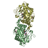 8a8iC 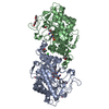 8au8C 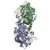 8au9C  8auaC  8aueC 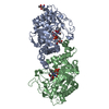 8aufC 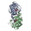 8augC 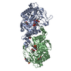 8auhC 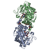 8auiC  8aujC  8aulC  8aumC  8aunC  8auoC  8auqC  3hgsS S: Starting model for refinement C: citing same article ( |
|---|---|
| Similar structure data |
-Links
 PDBj
PDBj- Assembly
| Deposited unit | 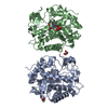
| ||||||||||||
|---|---|---|---|---|---|---|---|---|---|---|---|---|---|
| 1 | 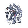
| ||||||||||||
| 2 | 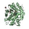
| ||||||||||||
| Unit cell |
|
-Components
| #1: Protein | Mass: 44401.203 Da / Num. of mol.: 2 Source method: isolated from a genetically manipulated source Source: (gene. exp.)  #2: Chemical |   Mass: 456.344 Da / Num. of mol.: 2 / Source method: obtained synthetically / Formula: C17H21N4O9P / Feature type: SUBJECT OF INVESTIGATION Mass: 456.344 Da / Num. of mol.: 2 / Source method: obtained synthetically / Formula: C17H21N4O9P / Feature type: SUBJECT OF INVESTIGATION#3: Chemical | 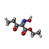  Mass: 173.167 Da / Num. of mol.: 2 / Source method: obtained synthetically / Formula: C7H11NO4 / Feature type: SUBJECT OF INVESTIGATION Mass: 173.167 Da / Num. of mol.: 2 / Source method: obtained synthetically / Formula: C7H11NO4 / Feature type: SUBJECT OF INVESTIGATION#4: Chemical | ChemComp-PEG /   Mass: 106.120 Da / Num. of mol.: 5 / Source method: obtained synthetically / Formula: C4H10O3 Mass: 106.120 Da / Num. of mol.: 5 / Source method: obtained synthetically / Formula: C4H10O3#5: Water | ChemComp-HOH / |  Mass: 18.015 Da / Num. of mol.: 403 / Source method: isolated from a natural source / Formula: H2O Mass: 18.015 Da / Num. of mol.: 403 / Source method: isolated from a natural source / Formula: H2OHas ligand of interest | Y | |
|---|
-Experimental details
-Experiment
| Experiment | Method: X-RAY DIFFRACTION / Number of used crystals: 1 |
|---|
- Sample preparation
| Crystal | Density Matthews: 2.2 Å3/Da / Density % sol: 43.97 % |
|---|---|
| Crystal grow | Temperature: 293.15 K / Method: vapor diffusion, sitting drop / pH: 8.5 / Details: 100 mM CaCl2,10 % w/v PEG 8000, 100 mM Tris pH 8.5 |
-Data collection
| Diffraction | Mean temperature: 100 K / Serial crystal experiment: N |
|---|---|
| Diffraction source | Source: SYNCHROTRON / Site: ESRF  / Beamline: MASSIF-1 / Wavelength: 0.965459 Å / Beamline: MASSIF-1 / Wavelength: 0.965459 Å |
| Detector | Type: DECTRIS PILATUS3 2M / Detector: PIXEL / Date: Apr 17, 2021 |
| Radiation | Protocol: SINGLE WAVELENGTH / Monochromatic (M) / Laue (L): M / Scattering type: x-ray |
| Radiation wavelength | Wavelength: 0.965459 Å / Relative weight: 1 |
| Reflection | Resolution: 1.62→44.2 Å / Num. obs: 176405 / % possible obs: 91.63 % / Redundancy: 1.6 % / Biso Wilson estimate: 17.69 Å2 / CC1/2: 0.987 / Net I/σ(I): 5.06 |
| Reflection shell | Resolution: 1.62→1.678 Å / Num. unique obs: 18096 / CC1/2: 0.619 |
- Processing
| Software |
| ||||||||||||||||||||||||||||||||||||||||||||||||||||||||||||||||||||||||||||||||||||||||||||||||||
|---|---|---|---|---|---|---|---|---|---|---|---|---|---|---|---|---|---|---|---|---|---|---|---|---|---|---|---|---|---|---|---|---|---|---|---|---|---|---|---|---|---|---|---|---|---|---|---|---|---|---|---|---|---|---|---|---|---|---|---|---|---|---|---|---|---|---|---|---|---|---|---|---|---|---|---|---|---|---|---|---|---|---|---|---|---|---|---|---|---|---|---|---|---|---|---|---|---|---|---|
| Refinement | Method to determine structure: MOLECULAR REPLACEMENT Starting model: 3HGS Resolution: 1.62→44.2 Å / SU ML: 0.1965 / Cross valid method: FREE R-VALUE / σ(F): 0.92 / Phase error: 22.8987 Stereochemistry target values: GeoStd + Monomer Library + CDL v1.2
| ||||||||||||||||||||||||||||||||||||||||||||||||||||||||||||||||||||||||||||||||||||||||||||||||||
| Solvent computation | Shrinkage radii: 0.9 Å / VDW probe radii: 1.11 Å / Solvent model: FLAT BULK SOLVENT MODEL | ||||||||||||||||||||||||||||||||||||||||||||||||||||||||||||||||||||||||||||||||||||||||||||||||||
| Displacement parameters | Biso mean: 21.33 Å2 | ||||||||||||||||||||||||||||||||||||||||||||||||||||||||||||||||||||||||||||||||||||||||||||||||||
| Refinement step | Cycle: LAST / Resolution: 1.62→44.2 Å
| ||||||||||||||||||||||||||||||||||||||||||||||||||||||||||||||||||||||||||||||||||||||||||||||||||
| Refine LS restraints |
| ||||||||||||||||||||||||||||||||||||||||||||||||||||||||||||||||||||||||||||||||||||||||||||||||||
| LS refinement shell |
|
