 Movie
Movie Controller


+ Open data

- Basic information
| Entry | Database: PDB / ID: 8ask | ||||||
|---|---|---|---|---|---|---|---|
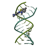
| Title | Crystal structure of d(GCCCACCACGGC) | ||||||
 Components Components |
| ||||||
 Keywords Keywords | DNA / Ligand / Interaction | ||||||
| Function / homology | DNA / DNA (> 10) Function and homology information Function and homology information | ||||||
| Biological species | DNA molecule (others) | ||||||
| Method |  X-RAY DIFFRACTION / SYNCHROTRON / MOLECULAR REPLACEMENT / Resolution: 2.955 Å X-RAY DIFFRACTION / SYNCHROTRON / MOLECULAR REPLACEMENT / Resolution: 2.955 Å | ||||||
 Authors Authors | Sbirkova-Dimitrova, H.I. / Shivachev, B.L. / Rusev, R. / Heroux, A. / Doukov, T. / Kuvandjiev, N. | ||||||
| Funding support | Bulgaria, 1items
| ||||||
 Citation Citation | Journal: Crystals / Year: 2022 Title: Structural Characterization of Alzheimer DNA Promoter Sequences from the Amyloid Precursor Gene in the Presence of Thioflavin T and Analogs Authors: Sbirkova-Dimitrova, H. / Rusew, R. / Kuvandjiev, N. / Heroux, A. / Doukov, T. / Shivachev, B.L. | ||||||
| History |
|
- Structure visualization
| Structure viewer | Molecule: MolmilJmol/JSmol |
|---|
- Downloads & links
-Download
| PDBx/mmCIF format | 8ask.cif.gz | 24.6 KB | Display | PDBx/mmCIF format |
|---|---|---|---|---|
| PDB format | pdb8ask.ent.gz | Display | PDB format | |
| PDBx/mmJSON format | 8ask.json.gz | Tree view | PDBx/mmJSON format | |
| Others |  Other downloads Other downloads |
-Validation report
| Summary document | 8ask_validation.pdf.gz | 376.9 KB | Display | wwPDB validaton report |
|---|---|---|---|---|
| Full document | 8ask_full_validation.pdf.gz | 376.8 KB | Display | |
| Data in XML | 8ask_validation.xml.gz | 2.3 KB | Display | |
| Data in CIF | 8ask_validation.cif.gz | 2.7 KB | Display | |
| Arichive directory | https://data.pdbj.org/pub/pdb/validation_reports/as/8askftp://data.pdbj.org/pub/pdb/validation_reports/as/8ask | HTTPS FTP |
-Related structure data
-Links
 PDBj
PDBj


- Assembly
| Deposited unit | 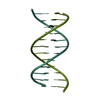
| ||||||||
|---|---|---|---|---|---|---|---|---|---|
| 1 |
| ||||||||
| Unit cell |
|
-Components
| #1: DNA chain | Mass: 3593.346 Da / Num. of mol.: 1 Source method: isolated from a genetically manipulated source Source: (gene. exp.) DNA molecule (others) / Production host:  Synthesium (invertebrata) Synthesium (invertebrata) |
|---|---|
| #2: DNA chain | Mass: 3735.414 Da / Num. of mol.: 1 Source method: isolated from a genetically manipulated source Source: (gene. exp.) DNA molecule (others) / Production host: Synthesium (invertebrata) |
-Experimental details
-Experiment
| Experiment | Method: X-RAY DIFFRACTION / Number of used crystals: 1 |
|---|
- Sample preparation
| Crystal | Density Matthews: 2.52 Å3/Da / Density % sol: 51.23 % |
|---|---|
| Crystal grow | Temperature: 294 K / Method: vapor diffusion, hanging drop / Details: 20%MPD, 120mMMgCl2, 60mM NaCaCo, 2mM Spermine |
-Data collection
| Diffraction | Mean temperature: 270 K / Serial crystal experiment: N |
|---|---|
| Diffraction source | Source: SYNCHROTRON / Site: ELETTRA  / Beamline: 5.2R / Wavelength: 2 Å / Beamline: 5.2R / Wavelength: 2 Å |
| Detector | Type: DECTRIS PILATUS 2M / Detector: PIXEL / Date: Aug 3, 2022 |
| Radiation | Protocol: SINGLE WAVELENGTH / Monochromatic (M) / Laue (L): M / Scattering type: x-ray |
| Radiation wavelength | Wavelength: 2 Å / Relative weight: 1 |
| Reflection | Resolution: 2.95→35.59 Å / Num. obs: 2783 / % possible obs: 96.95 % / Redundancy: 9.2 % / CC1/2: 0.992 / Net I/σ(I): 2 |
| Reflection shell | Resolution: 2.97→3.02 Å / Num. unique obs: 1399 / CC1/2: 0.3 |
- Processing
| Software |
| |||||||||||||||||||||||||||||||||||||||||||||||||||||||||||||||||||||||||||||||||||||||||||||||||||||||||||||||||||||||||||||||||||||||||||||||||||
|---|---|---|---|---|---|---|---|---|---|---|---|---|---|---|---|---|---|---|---|---|---|---|---|---|---|---|---|---|---|---|---|---|---|---|---|---|---|---|---|---|---|---|---|---|---|---|---|---|---|---|---|---|---|---|---|---|---|---|---|---|---|---|---|---|---|---|---|---|---|---|---|---|---|---|---|---|---|---|---|---|---|---|---|---|---|---|---|---|---|---|---|---|---|---|---|---|---|---|---|---|---|---|---|---|---|---|---|---|---|---|---|---|---|---|---|---|---|---|---|---|---|---|---|---|---|---|---|---|---|---|---|---|---|---|---|---|---|---|---|---|---|---|---|---|---|---|---|---|
| Refinement | Method to determine structure: MOLECULAR REPLACEMENT Starting model: X3DNA Resolution: 2.955→35.59 Å / Cor.coef. Fo:Fc: 0.987 / Cor.coef. Fo:Fc free: 0.891 / SU B: 27.017 / SU ML: 0.398 / Cross valid method: NONE / ESU R Free: 0.413 Details: Hydrogens have been added in their riding positions
| |||||||||||||||||||||||||||||||||||||||||||||||||||||||||||||||||||||||||||||||||||||||||||||||||||||||||||||||||||||||||||||||||||||||||||||||||||
| Solvent computation | Ion probe radii: 0.8 Å / Shrinkage radii: 0.8 Å / VDW probe radii: 1.2 Å / Solvent model: MASK BULK SOLVENT | |||||||||||||||||||||||||||||||||||||||||||||||||||||||||||||||||||||||||||||||||||||||||||||||||||||||||||||||||||||||||||||||||||||||||||||||||||
| Displacement parameters | Biso mean: 88.593 Å2
| |||||||||||||||||||||||||||||||||||||||||||||||||||||||||||||||||||||||||||||||||||||||||||||||||||||||||||||||||||||||||||||||||||||||||||||||||||
| Refinement step | Cycle: LAST / Resolution: 2.955→35.59 Å
| |||||||||||||||||||||||||||||||||||||||||||||||||||||||||||||||||||||||||||||||||||||||||||||||||||||||||||||||||||||||||||||||||||||||||||||||||||
| Refine LS restraints |
| |||||||||||||||||||||||||||||||||||||||||||||||||||||||||||||||||||||||||||||||||||||||||||||||||||||||||||||||||||||||||||||||||||||||||||||||||||
| LS refinement shell |
|
