 Movie
Movie Controller

[English] 日本語
 Yorodumi
- PDB-8aq6: NanoLuc luciferase with bound furimamide in surface allosteric site -
+ Open data

- Basic information
| Entry | Database: PDB / ID: 8aq6 | ||||||
|---|---|---|---|---|---|---|---|
| Title | NanoLuc luciferase with bound furimamide in surface allosteric site | ||||||
 Components Components | NanoLuc luciferase | ||||||
 Keywords Keywords | LUMINESCENT PROTEIN / Luciferase / NanoLuc / NLuc / luciferin / furimazine | ||||||
| Function / homology |  Function and homology information Function and homology informationOplophorus-luciferin 2-monooxygenase / Oplophorus-luciferin 2-monooxygenase activity / bioluminescence / extracellular region Similarity search - Function | ||||||
| Biological species |  Oplophorus gracilirostris (arthropod) Oplophorus gracilirostris (arthropod) | ||||||
| Method |  X-RAY DIFFRACTION / SYNCHROTRON / MOLECULAR REPLACEMENT / molecular replacement / Resolution: 1.69 Å X-RAY DIFFRACTION / SYNCHROTRON / MOLECULAR REPLACEMENT / molecular replacement / Resolution: 1.69 Å | ||||||
 Authors Authors | Nemergut, M. / Marek, M. | ||||||
| Funding support |  Czech Republic, 1items Czech Republic, 1items
| ||||||
 Citation Citation | Journal: Nat Commun / Year: 2023 Title: Illuminating the mechanism and allosteric behavior of NanoLuc luciferase. Authors: Nemergut, M. / Pluskal, D. / Horackova, J. / Sustrova, T. / Tulis, J. / Barta, T. / Baatallah, R. / Gagnot, G. / Novakova, V. / Majerova, M. / Sedlackova, K. / Marques, S.M. / Toul, M. / ...Authors: Nemergut, M. / Pluskal, D. / Horackova, J. / Sustrova, T. / Tulis, J. / Barta, T. / Baatallah, R. / Gagnot, G. / Novakova, V. / Majerova, M. / Sedlackova, K. / Marques, S.M. / Toul, M. / Damborsky, J. / Prokop, Z. / Bednar, D. / Janin, Y.L. / Marek, M. | ||||||
| History |
|
- Structure visualization
| Structure viewer | Molecule: MolmilJmol/JSmol |
|---|
- Downloads & links
-Download
| PDBx/mmCIF format | 8aq6.cif.gz | 595.4 KB | Display | PDBx/mmCIF format |
|---|---|---|---|---|
| PDB format | pdb8aq6.ent.gz | 489.8 KB | Display | PDB format |
| PDBx/mmJSON format | 8aq6.json.gz | Tree view | PDBx/mmJSON format | |
| Others |  Other downloads Other downloads |
-Validation report
| Arichive directory | https://data.pdbj.org/pub/pdb/validation_reports/aq/8aq6ftp://data.pdbj.org/pub/pdb/validation_reports/aq/8aq6 | HTTPS FTP |
|---|
-Related structure data
| Related structure data |  8aqhC 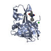 8aqiC 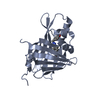 8bo9C 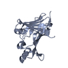 5b0uS S: Starting model for refinement C: citing same article ( |
|---|---|
| Similar structure data |
-Links
 PDBj
PDBj


- Assembly
| Deposited unit | 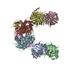
| ||||||||||||
|---|---|---|---|---|---|---|---|---|---|---|---|---|---|
| 1 | 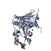
| ||||||||||||
| 2 | 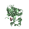
| ||||||||||||
| 3 | 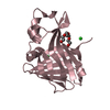
| ||||||||||||
| 4 | 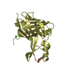
| ||||||||||||
| 5 | 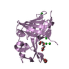
| ||||||||||||
| 6 | 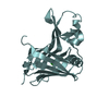
| ||||||||||||
| 7 | 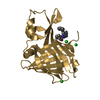
| ||||||||||||
| 8 | 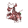
| ||||||||||||
| Unit cell |
| ||||||||||||
| Components on special symmetry positions |
|
-Components
-Protein , 1 types, 8 molecules ABCDEFGH
| #1: Protein | Mass: 20389.240 Da / Num. of mol.: 8 Source method: isolated from a genetically manipulated source Source: (gene. exp.) Oplophorus gracilirostris (arthropod)Production host:  References: UniProt: Q9GV45, Oplophorus-luciferin 2-monooxygenase |
|---|
-Non-polymers , 7 types, 804 molecules 


| #2: Chemical |  Mass: 92.094 Da / Num. of mol.: 2 / Source method: obtained synthetically / Formula: C3H8O3 / Feature type: SUBJECT OF INVESTIGATION Mass: 92.094 Da / Num. of mol.: 2 / Source method: obtained synthetically / Formula: C3H8O3 / Feature type: SUBJECT OF INVESTIGATION#3: Chemical | ChemComp-PG4 /  Mass: 194.226 Da / Num. of mol.: 7 / Source method: obtained synthetically / Formula: C8H18O5 / Feature type: SUBJECT OF INVESTIGATION / Comment: precipitant*YM Mass: 194.226 Da / Num. of mol.: 7 / Source method: obtained synthetically / Formula: C8H18O5 / Feature type: SUBJECT OF INVESTIGATION / Comment: precipitant*YM#4: Chemical | ChemComp-OXY / |  Mass: 31.999 Da / Num. of mol.: 1 / Source method: obtained synthetically / Formula: O2 / Feature type: SUBJECT OF INVESTIGATION Mass: 31.999 Da / Num. of mol.: 1 / Source method: obtained synthetically / Formula: O2 / Feature type: SUBJECT OF INVESTIGATION#5: Chemical | ChemComp-CL /  Mass: 35.453 Da / Num. of mol.: 22 / Source method: obtained synthetically / Formula: Cl / Feature type: SUBJECT OF INVESTIGATION Mass: 35.453 Da / Num. of mol.: 22 / Source method: obtained synthetically / Formula: Cl / Feature type: SUBJECT OF INVESTIGATION#6: Chemical | ChemComp-PGE / |  Mass: 150.173 Da / Num. of mol.: 1 / Source method: obtained synthetically / Formula: C6H14O4 / Feature type: SUBJECT OF INVESTIGATION Mass: 150.173 Da / Num. of mol.: 1 / Source method: obtained synthetically / Formula: C6H14O4 / Feature type: SUBJECT OF INVESTIGATION#7: Chemical | ChemComp-NT0 / |  Mass: 369.416 Da / Num. of mol.: 1 / Source method: obtained synthetically / Formula: C23H19N3O2 / Feature type: SUBJECT OF INVESTIGATION Mass: 369.416 Da / Num. of mol.: 1 / Source method: obtained synthetically / Formula: C23H19N3O2 / Feature type: SUBJECT OF INVESTIGATION#8: Water | ChemComp-HOH / | Mass: 18.015 Da / Num. of mol.: 770 / Source method: isolated from a natural source / Formula: H2O |
|---|
-Details
| Has ligand of interest | Y |
|---|---|
| Has protein modification | N |
-Experimental details
-Experiment
| Experiment | Method: X-RAY DIFFRACTION / Number of used crystals: 1 |
|---|
- Sample preparation
| Crystal | Density Matthews: 2.23 Å3/Da / Density % sol: 44.96 % |
|---|---|
| Crystal grow | Temperature: 293.15 K / Method: vapor diffusion, hanging drop / pH: 4 Details: 200 mM MgCl2, 100 mM KCl, 25 mM Na acetate pH = 4.0, PEG 400 |
-Data collection
| Diffraction | Mean temperature: 100 K / Serial crystal experiment: N | |||||||||||||||||||||||||
|---|---|---|---|---|---|---|---|---|---|---|---|---|---|---|---|---|---|---|---|---|---|---|---|---|---|---|
| Diffraction source | Source: SYNCHROTRON / Site: SLS  / Beamline: X06DA / Wavelength: 0.9999 Å / Beamline: X06DA / Wavelength: 0.9999 Å | |||||||||||||||||||||||||
| Detector | Type: DECTRIS PILATUS 2M-F / Detector: PIXEL / Date: Jul 14, 2021 | |||||||||||||||||||||||||
| Radiation | Protocol: SINGLE WAVELENGTH / Monochromatic (M) / Laue (L): M / Scattering type: x-ray | |||||||||||||||||||||||||
| Radiation wavelength | Wavelength: 0.9999 Å / Relative weight: 1 | |||||||||||||||||||||||||
| Reflection twin |
| |||||||||||||||||||||||||
| Reflection | Resolution: 1.69→47.94 Å / Num. obs: 158483 / % possible obs: 99.3 % / Redundancy: 3.4 % / CC1/2: 0.996 / Rmerge(I) obs: 0.066 / Net I/σ(I): 9.7 | |||||||||||||||||||||||||
| Reflection shell | Resolution: 1.69→1.73 Å / Num. unique obs: 14059 / CC1/2: 0.503 |
-Phasing
| Phasing | Method: molecular replacement |
|---|
- Processing
| Software |
| |||||||||||||||||||||||||||||||||||||||||||||||||||||||||||||||||
|---|---|---|---|---|---|---|---|---|---|---|---|---|---|---|---|---|---|---|---|---|---|---|---|---|---|---|---|---|---|---|---|---|---|---|---|---|---|---|---|---|---|---|---|---|---|---|---|---|---|---|---|---|---|---|---|---|---|---|---|---|---|---|---|---|---|---|
| Refinement | Method to determine structure: MOLECULAR REPLACEMENT Starting model: 5B0U Resolution: 1.69→47.94 Å / Cor.coef. Fo:Fc: 0.978 / Cor.coef. Fo:Fc free: 0.964 / SU B: 3.232 / SU ML: 0.052 / SU R Cruickshank DPI: 0.0258 / Cross valid method: THROUGHOUT / σ(F): 0 / ESU R: 0.026 / ESU R Free: 0.019 / Stereochemistry target values: MAXIMUM LIKELIHOOD Details: HYDROGENS HAVE BEEN ADDED IN THE RIDING POSITIONS U VALUES : REFINED INDIVIDUALLY
| |||||||||||||||||||||||||||||||||||||||||||||||||||||||||||||||||
| Solvent computation | Ion probe radii: 0.8 Å / Shrinkage radii: 0.8 Å / VDW probe radii: 1.2 Å / Solvent model: MASK | |||||||||||||||||||||||||||||||||||||||||||||||||||||||||||||||||
| Displacement parameters | Biso max: 169.07 Å2 / Biso mean: 31.334 Å2 / Biso min: 13.96 Å2
| |||||||||||||||||||||||||||||||||||||||||||||||||||||||||||||||||
| Refinement step | Cycle: final / Resolution: 1.69→47.94 Å
| |||||||||||||||||||||||||||||||||||||||||||||||||||||||||||||||||
| Refine LS restraints |
| |||||||||||||||||||||||||||||||||||||||||||||||||||||||||||||||||
| LS refinement shell | Resolution: 1.69→1.733 Å / Rfactor Rfree error: 0
|
