 Movie
Movie Controller


+ Open data

- Basic information
| Entry | Database: PDB / ID: 8app | |||||||||
|---|---|---|---|---|---|---|---|---|---|---|
| Title | AbLys1 endolysin from Acinetobacter baumannii phage AbTZA1 | |||||||||
 Components Components | Endolysin | |||||||||
 Keywords Keywords | HYDROLASE / Endolysin / Antibacterial / Enzybiotic | |||||||||
| Function / homology |  Function and homology information Function and homology informationviral release from host cell by cytolysis / peptidoglycan catabolic process / cell wall macromolecule catabolic process / lysozyme / lysozyme activity / host cell cytoplasm / defense response to bacterium Similarity search - Function | |||||||||
| Biological species |  Acinetobacter phage AbTZA1 (virus) Acinetobacter phage AbTZA1 (virus) | |||||||||
| Method |  X-RAY DIFFRACTION / SYNCHROTRON / MOLECULAR REPLACEMENT / Resolution: 1.82 Å X-RAY DIFFRACTION / SYNCHROTRON / MOLECULAR REPLACEMENT / Resolution: 1.82 Å | |||||||||
 Authors Authors | Premetis, G.E. / Stathi, A. / Papageorgiou, A.C. / Labrou, N.E. | |||||||||
| Funding support |  Greece, European Union, 2items Greece, European Union, 2items
| |||||||||
 Citation Citation | Journal: Febs J. / Year: 2023 Title: Characterization of a glycoside hydrolase endolysin from Acinetobacter baumannii phage AbTZA1 with high antibacterial potency and novel structural features. Authors: Premetis, G.E. / Stathi, A. / Papageorgiou, A.C. / Labrou, N.E. | |||||||||
| History |
|
- Structure visualization
| Structure viewer | Molecule: MolmilJmol/JSmol |
|---|
- Downloads & links
-Download
| PDBx/mmCIF format | 8app.cif.gz | 208.2 KB | Display | PDBx/mmCIF format |
|---|---|---|---|---|
| PDB format | pdb8app.ent.gz | 133.7 KB | Display | PDB format |
| PDBx/mmJSON format | 8app.json.gz | Tree view | PDBx/mmJSON format | |
| Others |  Other downloads Other downloads |
-Validation report
| Summary document | 8app_validation.pdf.gz | 469.9 KB | Display | wwPDB validaton report |
|---|---|---|---|---|
| Full document | 8app_full_validation.pdf.gz | 476.5 KB | Display | |
| Data in XML | 8app_validation.xml.gz | 34.5 KB | Display | |
| Data in CIF | 8app_validation.cif.gz | 49.9 KB | Display | |
| Arichive directory | https://data.pdbj.org/pub/pdb/validation_reports/ap/8appftp://data.pdbj.org/pub/pdb/validation_reports/ap/8app | HTTPS FTP |
-Related structure data
| Similar structure data |
|---|
-Links
 PDBj
PDBj


- Assembly
| Deposited unit | 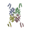
| ||||||||||||
|---|---|---|---|---|---|---|---|---|---|---|---|---|---|
| 1 | 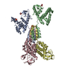
| ||||||||||||
| Unit cell |
|
-Components
| #1: Protein | Mass: 21642.762 Da / Num. of mol.: 4 Source method: isolated from a genetically manipulated source Source: (gene. exp.) Acinetobacter phage AbTZA1 (virus) / Plasmid: pETite-AbLys1-6His / Production host:  #2: Chemical | ChemComp-PO4 /   Mass: 94.971 Da / Num. of mol.: 4 / Source method: isolated from a natural source / Formula: PO4 Mass: 94.971 Da / Num. of mol.: 4 / Source method: isolated from a natural source / Formula: PO4#3: Chemical | ChemComp-GOL / |   Mass: 92.094 Da / Num. of mol.: 1 / Source method: obtained synthetically / Formula: C3H8O3 Mass: 92.094 Da / Num. of mol.: 1 / Source method: obtained synthetically / Formula: C3H8O3#4: Water | ChemComp-HOH / |  Mass: 18.015 Da / Num. of mol.: 587 / Source method: isolated from a natural source / Formula: H2O Mass: 18.015 Da / Num. of mol.: 587 / Source method: isolated from a natural source / Formula: H2OHas ligand of interest | N | |
|---|
-Experimental details
-Experiment
| Experiment | Method: X-RAY DIFFRACTION / Number of used crystals: 1 |
|---|
- Sample preparation
| Crystal | Density Matthews: 2.41 Å3/Da / Density % sol: 48.92 % / Description: Small rod-like crystals |
|---|---|
| Crystal grow | Temperature: 289 K / Method: vapor diffusion, hanging drop / pH: 4.2 Details: 0.1 M Phosphate/citrate, 0.2 M lithium sulphate, pH 4.2, 20% w/v PEG 1000 |
-Data collection
| Diffraction | Mean temperature: 100 K / Serial crystal experiment: N |
|---|---|
| Diffraction source | Source: SYNCHROTRON / Site: PETRA III, EMBL c/o DESY  / Beamline: P13 (MX1) / Wavelength: 0.8266 Å / Beamline: P13 (MX1) / Wavelength: 0.8266 Å |
| Detector | Type: DECTRIS EIGER X 16M / Detector: PIXEL / Date: Mar 3, 2022 / Details: KB mirrors |
| Radiation | Protocol: SINGLE WAVELENGTH / Monochromatic (M) / Laue (L): M / Scattering type: x-ray |
| Radiation wavelength | Wavelength: 0.8266 Å / Relative weight: 1 |
| Reflection | Resolution: 1.82→46.73 Å / Num. obs: 70067 / % possible obs: 96.2 % / Redundancy: 6.6 % / Biso Wilson estimate: 34.5 Å2 / CC1/2: 0.999 / Rpim(I) all: 0.042 / Rrim(I) all: 0.078 / Net I/σ(I): 12.5 |
| Reflection shell | Resolution: 1.82→1.88 Å / Redundancy: 3.2 % / Num. unique obs: 4406 / CC1/2: 0.424 / Rpim(I) all: 0.894 / Rrim(I) all: 1.276 / % possible all: 61.6 |
- Processing
| Software |
| |||||||||||||||||||||||||||||||||||||||||||||||||||||||||||||||||||||||||||||||||||||||||||||||||||||||||
|---|---|---|---|---|---|---|---|---|---|---|---|---|---|---|---|---|---|---|---|---|---|---|---|---|---|---|---|---|---|---|---|---|---|---|---|---|---|---|---|---|---|---|---|---|---|---|---|---|---|---|---|---|---|---|---|---|---|---|---|---|---|---|---|---|---|---|---|---|---|---|---|---|---|---|---|---|---|---|---|---|---|---|---|---|---|---|---|---|---|---|---|---|---|---|---|---|---|---|---|---|---|---|---|---|---|---|
| Refinement | Method to determine structure: MOLECULAR REPLACEMENT Starting model: AlphaFold model Resolution: 1.82→40.68 Å / SU ML: 0.25 / Cross valid method: FREE R-VALUE / σ(F): 1.34 / Phase error: 31.3615 Stereochemistry target values: GeoStd + Monomer Library + CDL v1.2
| |||||||||||||||||||||||||||||||||||||||||||||||||||||||||||||||||||||||||||||||||||||||||||||||||||||||||
| Solvent computation | Shrinkage radii: 0.9 Å / VDW probe radii: 1.1 Å / Solvent model: FLAT BULK SOLVENT MODEL | |||||||||||||||||||||||||||||||||||||||||||||||||||||||||||||||||||||||||||||||||||||||||||||||||||||||||
| Displacement parameters | Biso mean: 39.61 Å2 | |||||||||||||||||||||||||||||||||||||||||||||||||||||||||||||||||||||||||||||||||||||||||||||||||||||||||
| Refinement step | Cycle: LAST / Resolution: 1.82→40.68 Å
| |||||||||||||||||||||||||||||||||||||||||||||||||||||||||||||||||||||||||||||||||||||||||||||||||||||||||
| Refine LS restraints |
| |||||||||||||||||||||||||||||||||||||||||||||||||||||||||||||||||||||||||||||||||||||||||||||||||||||||||
| LS refinement shell |
|
