 Movie
Movie Controller


+ Open data

- Basic information
| Entry | Database: PDB / ID: 8ap0 | ||||||
|---|---|---|---|---|---|---|---|
| Title | ForT Mutant T138V | ||||||
 Components Components | Beta-ribofuranosylaminobenzene 5'-phosphate synthase | ||||||
 Keywords Keywords | STRUCTURAL PROTEIN / Formycin Biosynthesis pathway protein C-nucleoside formation enzyme | ||||||
| Function / homology |  Function and homology information Function and homology information | ||||||
| Biological species |  Streptomyces kaniharaensis (bacteria) Streptomyces kaniharaensis (bacteria) | ||||||
| Method |  X-RAY DIFFRACTION / SYNCHROTRON / MOLECULAR REPLACEMENT / Resolution: 1.6 Å X-RAY DIFFRACTION / SYNCHROTRON / MOLECULAR REPLACEMENT / Resolution: 1.6 Å | ||||||
 Authors Authors | Li, W. / Naismith, J.H. | ||||||
| Funding support |  United Kingdom, 1items United Kingdom, 1items
| ||||||
 Citation Citation | Journal: Open Biology / Year: 2023 Title: Experimental and computational snapshots of C-C bond formation in a C-nucleoside synthase. Authors: Li, W. / Girt, G.C. / Radadiya, A. / Stewart, J.J.P. / Richards, N.G.J. / Naismith, J.H. | ||||||
| History |
|
- Structure visualization
| Structure viewer | Molecule: MolmilJmol/JSmol |
|---|
- Downloads & links
-Download
| PDBx/mmCIF format | 8ap0.cif.gz | 153 KB | Display | PDBx/mmCIF format |
|---|---|---|---|---|
| PDB format | pdb8ap0.ent.gz | Display | PDB format | |
| PDBx/mmJSON format | 8ap0.json.gz | Tree view | PDBx/mmJSON format | |
| Others |  Other downloads Other downloads |
-Validation report
| Arichive directory | https://data.pdbj.org/pub/pdb/validation_reports/ap/8ap0ftp://data.pdbj.org/pub/pdb/validation_reports/ap/8ap0 | HTTPS FTP |
|---|
-Related structure data
| Related structure data |  8aozC 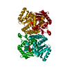 6yqqS S: Starting model for refinement C: citing same article ( |
|---|---|
| Similar structure data |
-Links
 PDBj
PDBj
- Assembly
| Deposited unit | 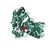
| ||||||||
|---|---|---|---|---|---|---|---|---|---|
| 1 | 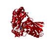
| ||||||||
| Unit cell |
|
-Components
-Protein / Sugars , 2 types, 2 molecules AAA
| #1: Protein | Mass: 36625.352 Da / Num. of mol.: 1 / Mutation: T138V Source method: isolated from a genetically manipulated source Details: Genetically mutated T138V / Source: (gene. exp.) Streptomyces kaniharaensis (bacteria) / Gene: cof6, F7Q99_03185 / Plasmid: pHis-TEV / Production host: |
|---|---|
| #2: Sugar | ChemComp-PRP /  Type: D-saccharide / Mass: 390.070 Da / Num. of mol.: 1 / Source method: obtained synthetically / Formula: C5H13O14P3 Type: D-saccharide / Mass: 390.070 Da / Num. of mol.: 1 / Source method: obtained synthetically / Formula: C5H13O14P3 |
-Non-polymers , 5 types, 315 molecules 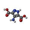


| #3: Chemical | ChemComp-MYO /  Mass: 171.111 Da / Num. of mol.: 1 / Source method: obtained synthetically / Formula: C5H5N3O4 / Feature type: SUBJECT OF INVESTIGATION Mass: 171.111 Da / Num. of mol.: 1 / Source method: obtained synthetically / Formula: C5H5N3O4 / Feature type: SUBJECT OF INVESTIGATION | ||||||
|---|---|---|---|---|---|---|---|
| #4: Chemical |  Mass: 92.094 Da / Num. of mol.: 2 / Source method: obtained synthetically / Formula: C3H8O3 Mass: 92.094 Da / Num. of mol.: 2 / Source method: obtained synthetically / Formula: C3H8O3#5: Chemical | ChemComp-K / |  Mass: 39.098 Da / Num. of mol.: 1 / Source method: obtained synthetically / Formula: K Mass: 39.098 Da / Num. of mol.: 1 / Source method: obtained synthetically / Formula: K#6: Chemical | ChemComp-MG / |  Mass: 24.305 Da / Num. of mol.: 1 / Source method: obtained synthetically / Formula: Mg Mass: 24.305 Da / Num. of mol.: 1 / Source method: obtained synthetically / Formula: Mg#7: Water | ChemComp-HOH / | Mass: 18.015 Da / Num. of mol.: 310 / Source method: isolated from a natural source / Formula: H2O |
-Details
| Has ligand of interest | Y |
|---|
-Experimental details
-Experiment
| Experiment | Method: X-RAY DIFFRACTION / Number of used crystals: 1 |
|---|
- Sample preparation
| Crystal | Density Matthews: 3.45 Å3/Da / Density % sol: 54.37 % / Description: Long needle shape crystal |
|---|---|
| Crystal grow | Temperature: 293.15 K / Method: vapor diffusion, hanging drop / pH: 7 Details: Bis-Tris Propane [20mM] pH7.0, Glycerol [20%], PEG 8000 [16%], Monopotassium phosphate [40mM] Temp details: Room Temperature |
-Data collection
| Diffraction | Mean temperature: 100 K / Ambient temp details: Cooled by liquid Nitrogen / Serial crystal experiment: N |
|---|---|
| Diffraction source | Source: SYNCHROTRON / Site: Diamond / Beamline: I04 / Wavelength: 0.9795 Å |
| Detector | Type: DECTRIS EIGER2 XE 16M / Detector: PIXEL / Date: Feb 17, 2021 |
| Radiation | Protocol: SINGLE WAVELENGTH / Monochromatic (M) / Laue (L): M / Scattering type: x-ray |
| Radiation wavelength | Wavelength: 0.9795 Å / Relative weight: 1 |
| Reflection | Resolution: 1.6→60 Å / Num. obs: 63680 / % possible obs: 100 % / Redundancy: 10.3 % / CC1/2: 1 / Net I/σ(I): 15 |
| Reflection shell | Resolution: 1.6→1.63 Å / Num. unique obs: 4624 / CC1/2: 0.8 |
- Processing
| Software |
| ||||||||||||||||||||||||||||||||||||||||||||||||||||||||||||||||||||||||||||||||||||||||||||||||||||||||||||||||||||||||||||||||||||||||||||||||||||||||||||||||
|---|---|---|---|---|---|---|---|---|---|---|---|---|---|---|---|---|---|---|---|---|---|---|---|---|---|---|---|---|---|---|---|---|---|---|---|---|---|---|---|---|---|---|---|---|---|---|---|---|---|---|---|---|---|---|---|---|---|---|---|---|---|---|---|---|---|---|---|---|---|---|---|---|---|---|---|---|---|---|---|---|---|---|---|---|---|---|---|---|---|---|---|---|---|---|---|---|---|---|---|---|---|---|---|---|---|---|---|---|---|---|---|---|---|---|---|---|---|---|---|---|---|---|---|---|---|---|---|---|---|---|---|---|---|---|---|---|---|---|---|---|---|---|---|---|---|---|---|---|---|---|---|---|---|---|---|---|---|---|---|---|---|
| Refinement | Method to determine structure: MOLECULAR REPLACEMENT Starting model: 6YQQ Resolution: 1.6→59.865 Å / Cor.coef. Fo:Fc: 0.981 / Cor.coef. Fo:Fc free: 0.969 / SU B: 6.61 / SU ML: 0.085 / Cross valid method: FREE R-VALUE / ESU R: 0.071 / ESU R Free: 0.071 Details: Hydrogens have been added in their riding positions
| ||||||||||||||||||||||||||||||||||||||||||||||||||||||||||||||||||||||||||||||||||||||||||||||||||||||||||||||||||||||||||||||||||||||||||||||||||||||||||||||||
| Solvent computation | Ion probe radii: 0.7 Å / Shrinkage radii: 0.7 Å / VDW probe radii: 1.3 Å / Solvent model: MASK BULK SOLVENT | ||||||||||||||||||||||||||||||||||||||||||||||||||||||||||||||||||||||||||||||||||||||||||||||||||||||||||||||||||||||||||||||||||||||||||||||||||||||||||||||||
| Displacement parameters | Biso mean: 31.414 Å2
| ||||||||||||||||||||||||||||||||||||||||||||||||||||||||||||||||||||||||||||||||||||||||||||||||||||||||||||||||||||||||||||||||||||||||||||||||||||||||||||||||
| Refinement step | Cycle: LAST / Resolution: 1.6→59.865 Å
| ||||||||||||||||||||||||||||||||||||||||||||||||||||||||||||||||||||||||||||||||||||||||||||||||||||||||||||||||||||||||||||||||||||||||||||||||||||||||||||||||
| Refine LS restraints |
| ||||||||||||||||||||||||||||||||||||||||||||||||||||||||||||||||||||||||||||||||||||||||||||||||||||||||||||||||||||||||||||||||||||||||||||||||||||||||||||||||
| LS refinement shell |
|
