 Movie
Movie Controller

[English] 日本語
 Yorodumi
- PDB-8ang: Structure of the amyloid-forming peptide LYIQWL from Tc5b, grown ... -
+ Open data

- Basic information
| Entry | Database: PDB / ID: 8ang | ||||||||||||
|---|---|---|---|---|---|---|---|---|---|---|---|---|---|
| Title | Structure of the amyloid-forming peptide LYIQWL from Tc5b, grown from 30% ethanol | ||||||||||||
 Components Components | Peptide LYIQWL from Tc5b | ||||||||||||
 Keywords Keywords | PROTEIN FIBRIL / amyloid | ||||||||||||
| Function / homology | ETHANOL Function and homology information Function and homology information | ||||||||||||
| Biological species | synthetic construct (others) | ||||||||||||
| Method |  X-RAY DIFFRACTION / MOLECULAR REPLACEMENT / Resolution: 1.5 Å X-RAY DIFFRACTION / MOLECULAR REPLACEMENT / Resolution: 1.5 Å | ||||||||||||
 Authors Authors | Durvanger, Z. | ||||||||||||
| Funding support |  Hungary, European Union, 3items Hungary, European Union, 3items
| ||||||||||||
 Citation Citation | Journal: Nat Commun / Year: 2023 Title: Polymorphic amyloid nanostructures of hormone peptides involved in glucose homeostasis display reversible amyloid formation. Authors: Horvath, D. / Durvanger, Z. / K Menyhard, D. / Sulyok-Eiler, M. / Bencs, F. / Gyulai, G. / Horvath, P. / Taricska, N. / Perczel, A. | ||||||||||||
| History |
|
- Structure visualization
| Structure viewer | Molecule: MolmilJmol/JSmol |
|---|
- Downloads & links
-Download
| PDBx/mmCIF format | 8ang.cif.gz | 13.9 KB | Display | PDBx/mmCIF format |
|---|---|---|---|---|
| PDB format | pdb8ang.ent.gz | 6.3 KB | Display | PDB format |
| PDBx/mmJSON format | 8ang.json.gz | Tree view | PDBx/mmJSON format | |
| Others |  Other downloads Other downloads |
-Validation report
| Arichive directory | https://data.pdbj.org/pub/pdb/validation_reports/an/8angftp://data.pdbj.org/pub/pdb/validation_reports/an/8ang | HTTPS FTP |
|---|
-Related structure data
| Related structure data | 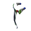 8anhC  8aniC 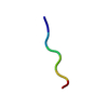 8anjC  8ankC  8anlC  8anmC 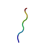 8annC 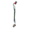 8onqC C: citing same article ( |
|---|---|
| Similar structure data |
-Links
 PDBj
PDBj


- Assembly
| Deposited unit | 
| ||||||||||||
|---|---|---|---|---|---|---|---|---|---|---|---|---|---|
| 1 |
| ||||||||||||
| Unit cell |
|
-Components
| #1: Protein/peptide | Mass: 835.001 Da / Num. of mol.: 1 / Source method: obtained synthetically / Source: (synth.) synthetic construct (others) |
|---|---|
| #2: Chemical | ChemComp-EOH /   Mass: 46.068 Da / Num. of mol.: 1 / Source method: obtained synthetically / Formula: C2H6O Mass: 46.068 Da / Num. of mol.: 1 / Source method: obtained synthetically / Formula: C2H6O |
| Has ligand of interest | N |
| Has protein modification | N |
-Experimental details
-Experiment
| Experiment | Method: X-RAY DIFFRACTION / Number of used crystals: 1 |
|---|
- Sample preparation
| Crystal | Density Matthews: 1.41 Å3/Da |
|---|---|
| Crystal grow | Temperature: 293 K / Method: evaporation, recrystallization Details: Peptide was dissolved in 30% ethanol at 0.5 mg/ml concentration, then incubated at 293K overnight. |
-Data collection
| Diffraction | Mean temperature: 100 K / Serial crystal experiment: N |
|---|---|
| Diffraction source | Source: ROTATING ANODE / Type: RIGAKU PhotonJet-R / Wavelength: 1.54184 Å |
| Detector | Type: RIGAKU HyPix-6000HE / Detector: PIXEL / Date: Jan 21, 2022 |
| Radiation | Protocol: SINGLE WAVELENGTH / Monochromatic (M) / Laue (L): M / Scattering type: x-ray |
| Radiation wavelength | Wavelength: 1.54184 Å / Relative weight: 1 |
| Reflection | Resolution: 1.5→20.5 Å / Num. obs: 895 / % possible obs: 95.01 % / Redundancy: 4.06 % / Biso Wilson estimate: 5.59 Å2 / CC1/2: 0.998 / Rrim(I) all: 0.066 / Net I/σ(I): 13.5 |
| Reflection shell | Resolution: 1.5→1.55 Å / Mean I/σ(I) obs: 3.8 / Num. unique obs: 61 / CC1/2: 0.978 / Rrim(I) all: 0.204 / % possible all: 65.59 |
- Processing
| Software |
| ||||||||||||||||||||||||
|---|---|---|---|---|---|---|---|---|---|---|---|---|---|---|---|---|---|---|---|---|---|---|---|---|---|
| Refinement | Method to determine structure: MOLECULAR REPLACEMENT Starting model: ideal 5 residue beta strand form the software Fragon Resolution: 1.5→20.5 Å / SU ML: -0 / Cross valid method: FREE R-VALUE / σ(F): 1.37 / Phase error: 4.4171 Stereochemistry target values: GeoStd + Monomer Library + CDL v1.2
| ||||||||||||||||||||||||
| Solvent computation | Shrinkage radii: 0.9 Å / VDW probe radii: 1.11 Å / Solvent model: FLAT BULK SOLVENT MODEL | ||||||||||||||||||||||||
| Displacement parameters | Biso mean: 4.33 Å2 | ||||||||||||||||||||||||
| Refinement step | Cycle: LAST / Resolution: 1.5→20.5 Å /
| ||||||||||||||||||||||||
| Refine LS restraints |
| ||||||||||||||||||||||||
| LS refinement shell | Resolution: 1.5→20.5 Å
|
