 Movie
Movie Controller


+ Open data

- Basic information
| Entry | Database: PDB / ID: 8ane | ||||||
|---|---|---|---|---|---|---|---|
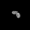
| Title | Structure of the type I-G CRISPR effector | ||||||
 Components Components |
| ||||||
 Keywords Keywords | IMMUNE SYSTEM / CRISPR effector | ||||||
| Function / homology | CRISPR-associated protein GSU0053 / CRISPR-associated protein GSU0053 (Cas_GSU0053) / RNA / RNA (> 10) / Type I-U CRISPR-associated protein Cas7 Function and homology information Function and homology information | ||||||
| Biological species |  Thioalkalivibrio sulfidiphilus HL-EbGr7 (bacteria) Thioalkalivibrio sulfidiphilus HL-EbGr7 (bacteria)synthetic construct (others) | ||||||
| Method | ELECTRON MICROSCOPY / single particle reconstruction / cryo EM / Resolution: 3.2 Å | ||||||
 Authors Authors | Shangguan, Q. / Graham, S. / Sundaramoorthy, R. / White, M.F. | ||||||
| Funding support |  United Kingdom, 1items United Kingdom, 1items
| ||||||
 Citation Citation | Journal: Nucleic Acids Res / Year: 2022 Title: Structure and mechanism of the type I-G CRISPR effector. Authors: Qilin Shangguan / Shirley Graham / Ramasubramanian Sundaramoorthy / Malcolm F White / Abstract: Type I CRISPR systems are the most common CRISPR type found in bacteria. They use a multisubunit effector, guided by crRNA, to detect and bind dsDNA targets, forming an R-loop and recruiting the Cas3 ...Type I CRISPR systems are the most common CRISPR type found in bacteria. They use a multisubunit effector, guided by crRNA, to detect and bind dsDNA targets, forming an R-loop and recruiting the Cas3 enzyme to facilitate target DNA destruction, thus providing immunity against mobile genetic elements. Subtypes have been classified into families A-G, with type I-G being the least well understood. Here, we report the composition, structure and function of the type I-G Cascade CRISPR effector from Thioalkalivibrio sulfidiphilus, revealing key new molecular details. The unique Csb2 subunit processes pre-crRNA, remaining bound to the 3' end of the mature crRNA, and seven Cas7 subunits form the backbone of the effector. Cas3 associates stably with the effector complex via the Cas8g subunit and is important for target DNA recognition. Structural analysis by cryo-Electron Microscopy reveals a strikingly curved backbone conformation with Cas8g spanning the belly of the structure. These biochemical and structural insights shed new light on the diversity of type I systems and open the way to applications in genome engineering. | ||||||
| History |
|
- Structure visualization
| Structure viewer | Molecule: MolmilJmol/JSmol |
|---|
- Downloads & links
-Download
| PDBx/mmCIF format | 8ane.cif.gz | 748.2 KB | Display | PDBx/mmCIF format |
|---|---|---|---|---|
| PDB format | pdb8ane.ent.gz | 632.6 KB | Display | PDB format |
| PDBx/mmJSON format | 8ane.json.gz | Tree view | PDBx/mmJSON format | |
| Others |  Other downloads Other downloads |
-Validation report
| Arichive directory | https://data.pdbj.org/pub/pdb/validation_reports/an/8aneftp://data.pdbj.org/pub/pdb/validation_reports/an/8ane | HTTPS FTP |
|---|
-Related structure data
| Related structure data | 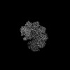 15540MC 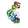 8b2xC M: map data used to model this data C: citing same article ( |
|---|---|
| Similar structure data |
-Links
 PDBj
PDBj


- Assembly
| Deposited unit | 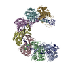
|
|---|---|
| 1 |
|
-Components
| #1: Protein | Mass: 34833.082 Da / Num. of mol.: 7 Source method: isolated from a genetically manipulated source Source: (gene. exp.) Thioalkalivibrio sulfidiphilus HL-EbGr7 (bacteria)Strain: HL-EbGR7 / Gene: Tgr7_2440 / Production host: #2: RNA chain | | Mass: 21082.465 Da / Num. of mol.: 1 / Source method: obtained synthetically / Source: (synth.) synthetic construct (others) |
|---|
-Experimental details
-Experiment
| Experiment | Method: ELECTRON MICROSCOPY |
|---|---|
| EM experiment | Aggregation state: PARTICLE / 3D reconstruction method: single particle reconstruction |
- Sample preparation
| Component | Name: Type IG cascade core cas7 with CrRNA / Type: COMPLEX / Entity ID: all / Source: RECOMBINANT |
|---|---|
| Molecular weight | Value: 0.035 MDa / Experimental value: NO |
| Source (natural) | Organism: Thioalkalivibrio sulfidiphilus HL-EbGr7 (bacteria) |
| Source (recombinant) | Organism: |
| Buffer solution | pH: 7.5 / Details: 20 mM Tris-HCl, 250 mM NaCl, pH 7.5 |
| Specimen | Conc.: 1 mg/ml / Embedding applied: NO / Shadowing applied: NO / Staining applied: NO / Vitrification applied: YES Details: Size exclusion purified monodisperse sample in solution |
| Specimen support | Grid material: COPPER / Grid mesh size: 400 divisions/in. / Grid type: Quantifoil R2/1 |
| Vitrification | Instrument: FEI VITROBOT MARK IV / Cryogen name: ETHANE / Humidity: 100 % / Chamber temperature: 4 K / Details: blot time 3.5 blot force 4 |
- Electron microscopy imaging
| Experimental equipment |  Model: Titan Krios / Image courtesy: FEI Company |
|---|---|
| Microscopy | Model: FEI TITAN KRIOS |
| Electron gun | Electron source:  FIELD EMISSION GUN / Accelerating voltage: 300 kV / Illumination mode: OTHER FIELD EMISSION GUN / Accelerating voltage: 300 kV / Illumination mode: OTHER |
| Electron lens | Mode: BRIGHT FIELD / Nominal magnification: 105000 X / Calibrated magnification: 105000 X / Nominal defocus max: 2800 nm / Nominal defocus min: 1600 nm / Calibrated defocus min: 1600 nm / Calibrated defocus max: 2800 nm / Cs: 2 mm / C2 aperture diameter: 70 µm / Alignment procedure: ZEMLIN TABLEAU |
| Specimen holder | Cryogen: NITROGEN / Specimen holder model: FEI TITAN KRIOS AUTOGRID HOLDER / Temperature (max): 183 K / Temperature (min): 77 K |
| Image recording | Average exposure time: 3 sec. / Electron dose: 50 e/Å2 / Film or detector model: GATAN K3 BIOQUANTUM (6k x 4k) / Num. of grids imaged: 1 / Num. of real images: 15710 |
| EM imaging optics | Energyfilter name: GIF Bioquantum / Energyfilter slit width: 20 eV |
| Image scans | Sampling size: 0.84 µm / Width: 5000 / Height: 4000 |
- Processing
| Software | Name: PHENIX / Version: dev_4674: / Classification: refinement | ||||||||||||||||||||||||||||||||||||||||||||
|---|---|---|---|---|---|---|---|---|---|---|---|---|---|---|---|---|---|---|---|---|---|---|---|---|---|---|---|---|---|---|---|---|---|---|---|---|---|---|---|---|---|---|---|---|---|
| EM software |
| ||||||||||||||||||||||||||||||||||||||||||||
| CTF correction | Type: PHASE FLIPPING ONLY | ||||||||||||||||||||||||||||||||||||||||||||
| Particle selection | Num. of particles selected: 1000 | ||||||||||||||||||||||||||||||||||||||||||||
| 3D reconstruction | Resolution: 3.2 Å / Resolution method: FSC 0.143 CUT-OFF / Num. of particles: 415000 / Algorithm: FOURIER SPACE / Symmetry type: POINT | ||||||||||||||||||||||||||||||||||||||||||||
| Atomic model building | B value: 188 / Protocol: AB INITIO MODEL / Space: REAL / Target criteria: correlation coefficient | ||||||||||||||||||||||||||||||||||||||||||||
| Refine LS restraints |
|
