 Movie
Movie Controller


+ Open data

- Basic information
| Entry | Database: PDB / ID: 8an4 | ||||||
|---|---|---|---|---|---|---|---|
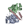
| Title | MenT1 toxin (rv0078a) from Mycobacterium tuberculosis H37Rv | ||||||
 Components Components | Bacterial toxin | ||||||
 Keywords Keywords | TOXIN / Toxin-antitoxin Tuberculosis Nucleotidyltransferase MenT MenAT | ||||||
| Function / homology | Beta Polymerase; domain 2 - #40 / Nucleotidyl transferase AbiEii toxin, Type IV TA system / Nucleotidyl transferase AbiEii toxin, Type IV TA system / Beta Polymerase; domain 2 / Nucleotidyltransferase superfamily / 2-Layer Sandwich / Alpha Beta / Nucleotidyl transferase AbiEii/AbiGii toxin family protein Function and homology information Function and homology information | ||||||
| Biological species |  Mycobacterium tuberculosis H37Rv (bacteria) Mycobacterium tuberculosis H37Rv (bacteria) | ||||||
| Method |  X-RAY DIFFRACTION / SYNCHROTRON / MOLECULAR REPLACEMENT / Resolution: 1.65 Å X-RAY DIFFRACTION / SYNCHROTRON / MOLECULAR REPLACEMENT / Resolution: 1.65 Å | ||||||
 Authors Authors | Xu, X. / Usher, B. / Gutierrez, C. / Barriot, R. / Arrowsmith, T.J. / Han, X. / Redder, P. / Neyrolles, O. / Blower, T.R. / Genevaux, P. | ||||||
| Funding support | 1items
| ||||||
 Citation Citation | Journal: Nat Commun / Year: 2023 Title: MenT nucleotidyltransferase toxins extend tRNA acceptor stems and can be inhibited by asymmetrical antitoxin binding. Authors: Xu, X. / Usher, B. / Gutierrez, C. / Barriot, R. / Arrowsmith, T.J. / Han, X. / Redder, P. / Neyrolles, O. / Blower, T.R. / Genevaux, P. | ||||||
| History |
|
- Structure visualization
| Structure viewer | Molecule: MolmilJmol/JSmol |
|---|
- Downloads & links
-Download
| PDBx/mmCIF format | 8an4.cif.gz | 230.7 KB | Display | PDBx/mmCIF format |
|---|---|---|---|---|
| PDB format | pdb8an4.ent.gz | 181.9 KB | Display | PDB format |
| PDBx/mmJSON format | 8an4.json.gz | Tree view | PDBx/mmJSON format | |
| Others |  Other downloads Other downloads |
-Validation report
| Arichive directory | https://data.pdbj.org/pub/pdb/validation_reports/an/8an4ftp://data.pdbj.org/pub/pdb/validation_reports/an/8an4 | HTTPS FTP |
|---|
-Related structure data
-Links
 PDBj
PDBj- Assembly
| Deposited unit | 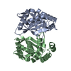
| ||||||||||||
|---|---|---|---|---|---|---|---|---|---|---|---|---|---|
| 1 | 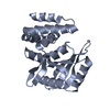
| ||||||||||||
| 2 | 
| ||||||||||||
| Unit cell |
|
-Components
| #1: Protein | Mass: 21509.369 Da / Num. of mol.: 2 Source method: isolated from a genetically manipulated source Source: (gene. exp.) Mycobacterium tuberculosis H37Rv (bacteria)Strain: ATCC 25618 / H37Rv / Gene: Rv0078A / Production host: #2: Water | ChemComp-HOH / |  Mass: 18.015 Da / Num. of mol.: 120 / Source method: isolated from a natural source / Formula: H2O Mass: 18.015 Da / Num. of mol.: 120 / Source method: isolated from a natural source / Formula: H2O |
|---|
-Experimental details
-Experiment
| Experiment | Method: X-RAY DIFFRACTION / Number of used crystals: 1 |
|---|
- Sample preparation
| Crystal | Density Matthews: 2.64 Å3/Da / Density % sol: 53.46 % |
|---|---|
| Crystal grow | Temperature: 294 K / Method: vapor diffusion, hanging drop / Details: 1.8 M lithium sulphate 0.1 M Tris pH 7.5 |
-Data collection
| Diffraction | Mean temperature: 100 K / Serial crystal experiment: N |
|---|---|
| Diffraction source | Source: SYNCHROTRON / Site: Diamond  / Beamline: I24 / Wavelength: 0.9795 Å / Beamline: I24 / Wavelength: 0.9795 Å |
| Detector | Type: DECTRIS PILATUS 6M / Detector: PIXEL / Date: Dec 16, 2020 |
| Radiation | Protocol: SINGLE WAVELENGTH / Monochromatic (M) / Laue (L): M / Scattering type: x-ray |
| Radiation wavelength | Wavelength: 0.9795 Å / Relative weight: 1 |
| Reflection | Resolution: 1.65→43.84 Å / Num. obs: 55309 / % possible obs: 90.68 % / Redundancy: 102.3 % / Biso Wilson estimate: 36.95 Å2 / CC1/2: 0.99 / Net I/σ(I): 8.95 |
| Reflection shell | Resolution: 1.65→1.71 Å / Num. unique obs: 5434 / CC1/2: 0.29 |
- Processing
| Software |
| ||||||||||||||||||||||||||||||||||||||||||||||||||||||||||||||||||||||||||||||||||||||||||||||||||||||||||||||||||||||||||||||
|---|---|---|---|---|---|---|---|---|---|---|---|---|---|---|---|---|---|---|---|---|---|---|---|---|---|---|---|---|---|---|---|---|---|---|---|---|---|---|---|---|---|---|---|---|---|---|---|---|---|---|---|---|---|---|---|---|---|---|---|---|---|---|---|---|---|---|---|---|---|---|---|---|---|---|---|---|---|---|---|---|---|---|---|---|---|---|---|---|---|---|---|---|---|---|---|---|---|---|---|---|---|---|---|---|---|---|---|---|---|---|---|---|---|---|---|---|---|---|---|---|---|---|---|---|---|---|---|
| Refinement | Method to determine structure: MOLECULAR REPLACEMENT Starting model: tbc Resolution: 1.65→43.84 Å / SU ML: 0.2888 / Cross valid method: FREE R-VALUE / σ(F): 1.33 / Phase error: 32.5608 Stereochemistry target values: GeoStd + Monomer Library + CDL v1.2
| ||||||||||||||||||||||||||||||||||||||||||||||||||||||||||||||||||||||||||||||||||||||||||||||||||||||||||||||||||||||||||||||
| Solvent computation | Shrinkage radii: 0.9 Å / VDW probe radii: 1.11 Å / Solvent model: FLAT BULK SOLVENT MODEL | ||||||||||||||||||||||||||||||||||||||||||||||||||||||||||||||||||||||||||||||||||||||||||||||||||||||||||||||||||||||||||||||
| Displacement parameters | Biso mean: 43.87 Å2 | ||||||||||||||||||||||||||||||||||||||||||||||||||||||||||||||||||||||||||||||||||||||||||||||||||||||||||||||||||||||||||||||
| Refinement step | Cycle: LAST / Resolution: 1.65→43.84 Å
| ||||||||||||||||||||||||||||||||||||||||||||||||||||||||||||||||||||||||||||||||||||||||||||||||||||||||||||||||||||||||||||||
| Refine LS restraints |
| ||||||||||||||||||||||||||||||||||||||||||||||||||||||||||||||||||||||||||||||||||||||||||||||||||||||||||||||||||||||||||||||
| LS refinement shell |
|
