 Movie
Movie Controller

[English] 日本語
 Yorodumi
- PDB-8aij: STRUCTURE OF THE LECB LECTIN FROM PSEUDOMONAS AERUGINOSA STRAIN P... -
+ Open data

- Basic information
| Entry | Database: PDB / ID: 8aij | ||||||
|---|---|---|---|---|---|---|---|
| Title | STRUCTURE OF THE LECB LECTIN FROM PSEUDOMONAS AERUGINOSA STRAIN PAO1 IN COMPLEX WITH N-(alpha-L-Fucopyranosyl)benzamide (6) | ||||||
 Components Components | Fucose-binding lectin PA-IIL | ||||||
 Keywords Keywords | SUGAR BINDING PROTEIN / P. aeruginosa lectin / LecB / Inhibitor | ||||||
| Function / homology | Lectin, sugar-binding / Calcium-mediated lectin / Calcium-mediated lectin superfamily / Fucose-binding lectin II (PA-IIL) / single-species biofilm formation / carbohydrate binding / metal ion binding / N-(alpha-L-Fucopyranosyl)benzamide / Fucose-binding lectin PA-IIL Function and homology information Function and homology information | ||||||
| Biological species |  Pseudomonas aeruginosa PAO1 (bacteria) Pseudomonas aeruginosa PAO1 (bacteria) | ||||||
| Method |  X-RAY DIFFRACTION / SYNCHROTRON / MOLECULAR REPLACEMENT / Resolution: 1.5 Å X-RAY DIFFRACTION / SYNCHROTRON / MOLECULAR REPLACEMENT / Resolution: 1.5 Å | ||||||
 Authors Authors | Meiers, J. / Mala, P. / Varrot, A. / Siebs, E. / Imberty, A. / Titz, A. | ||||||
| Funding support |  France, 1items France, 1items
| ||||||
 Citation Citation | Journal: J.Med.Chem. / Year: 2022 Title: Discovery of N -beta-l-Fucosyl Amides as High-Affinity Ligands for the Pseudomonas aeruginosa Lectin LecB. Authors: Mala, P. / Siebs, E. / Meiers, J. / Rox, K. / Varrot, A. / Imberty, A. / Titz, A. | ||||||
| History |
|
- Structure visualization
| Structure viewer | Molecule: MolmilJmol/JSmol |
|---|
- Downloads & links
-Download
| PDBx/mmCIF format | 8aij.cif.gz | 118.6 KB | Display | PDBx/mmCIF format |
|---|---|---|---|---|
| PDB format | pdb8aij.ent.gz | Display | PDB format | |
| PDBx/mmJSON format | 8aij.json.gz | Tree view | PDBx/mmJSON format | |
| Others |  Other downloads Other downloads |
-Validation report
| Arichive directory | https://data.pdbj.org/pub/pdb/validation_reports/ai/8aijftp://data.pdbj.org/pub/pdb/validation_reports/ai/8aij | HTTPS FTP |
|---|
-Related structure data
| Related structure data |  8aiyC  5a3oS S: Starting model for refinement C: citing same article ( |
|---|---|
| Similar structure data |
-Links
 PDBj
PDBj- Assembly
| Deposited unit | 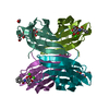
| ||||||||
|---|---|---|---|---|---|---|---|---|---|
| 1 |
| ||||||||
| Unit cell |
|
-Components
| #1: Protein | Mass: 11734.707 Da / Num. of mol.: 4 Source method: isolated from a genetically manipulated source Source: (gene. exp.) Pseudomonas aeruginosa PAO1 (bacteria) / Gene: lecB, PA3361 / Production host: #2: Chemical | ChemComp-CA /   Mass: 40.078 Da / Num. of mol.: 8 / Source method: obtained synthetically / Formula: Ca Mass: 40.078 Da / Num. of mol.: 8 / Source method: obtained synthetically / Formula: Ca#3: Chemical | ChemComp-M9I / 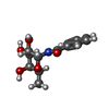  Mass: 267.278 Da / Num. of mol.: 4 / Source method: obtained synthetically / Formula: C13H17NO5 / Feature type: SUBJECT OF INVESTIGATION Mass: 267.278 Da / Num. of mol.: 4 / Source method: obtained synthetically / Formula: C13H17NO5 / Feature type: SUBJECT OF INVESTIGATION#4: Chemical | ChemComp-EDO /   Mass: 62.068 Da / Num. of mol.: 4 / Source method: obtained synthetically / Formula: C2H6O2 Mass: 62.068 Da / Num. of mol.: 4 / Source method: obtained synthetically / Formula: C2H6O2#5: Water | ChemComp-HOH / |  Mass: 18.015 Da / Num. of mol.: 509 / Source method: isolated from a natural source / Formula: H2O Mass: 18.015 Da / Num. of mol.: 509 / Source method: isolated from a natural source / Formula: H2OHas ligand of interest | Y | |
|---|
-Experimental details
-Experiment
| Experiment | Method: X-RAY DIFFRACTION / Number of used crystals: 1 |
|---|
- Sample preparation
| Crystal | Density Matthews: 2.21 Å3/Da / Density % sol: 44.44 % / Description: plates |
|---|---|
| Crystal grow | Temperature: 293.15 K / Method: vapor diffusion, hanging drop / pH: 4.4 Details: 24% PEG 8K, 1M LiCl and 100 mM sodium acetate pH = 4.4 |
-Data collection
| Diffraction | Mean temperature: 100 K / Serial crystal experiment: N |
|---|---|
| Diffraction source | Source: SYNCHROTRON / Site: SOLEIL / Beamline: PROXIMA 2 / Wavelength: 0.9801 Å |
| Detector | Type: DECTRIS EIGER X 9M / Detector: PIXEL / Date: Jun 19, 2022 Details: Cryogenically cooled channel cut crystal monochromator, a convex prefocussing mirror and a Kirkpatrick-Baez pair of focussing mirrors |
| Radiation | Protocol: SINGLE WAVELENGTH / Monochromatic (M) / Laue (L): M / Scattering type: x-ray |
| Radiation wavelength | Wavelength: 0.9801 Å / Relative weight: 1 |
| Reflection | Resolution: 1.5→40.69 Å / Num. obs: 211861 / % possible obs: 95.5 % / Redundancy: 3.4 % / CC1/2: 0.997 / Rmerge(I) obs: 0.051 / Rpim(I) all: 0.051 / Rrim(I) all: 0.072 / Net I/σ(I): 12.9 |
| Reflection shell | Resolution: 1.5→1.53 Å / Redundancy: 3.3 % / Rmerge(I) obs: 1.53 / Mean I/σ(I) obs: 3.4 / Num. unique obs: 9941 / CC1/2: 0.881 / Rpim(I) all: 0.318 / Rrim(I) all: 0.45 / % possible all: 92.49 |
- Processing
| Software |
| ||||||||||||||||||||||||||||||||||||||||||||||||||||||||||||||||||||||||||||||||||||||||||||||||||||||||||||||||||||||||||||||||||||||||||||||||||||||||||||||||
|---|---|---|---|---|---|---|---|---|---|---|---|---|---|---|---|---|---|---|---|---|---|---|---|---|---|---|---|---|---|---|---|---|---|---|---|---|---|---|---|---|---|---|---|---|---|---|---|---|---|---|---|---|---|---|---|---|---|---|---|---|---|---|---|---|---|---|---|---|---|---|---|---|---|---|---|---|---|---|---|---|---|---|---|---|---|---|---|---|---|---|---|---|---|---|---|---|---|---|---|---|---|---|---|---|---|---|---|---|---|---|---|---|---|---|---|---|---|---|---|---|---|---|---|---|---|---|---|---|---|---|---|---|---|---|---|---|---|---|---|---|---|---|---|---|---|---|---|---|---|---|---|---|---|---|---|---|---|---|---|---|---|
| Refinement | Method to determine structure: MOLECULAR REPLACEMENT Starting model: 5A3O Resolution: 1.5→40.69 Å / Cor.coef. Fo:Fc: 0.975 / Cor.coef. Fo:Fc free: 0.966 / SU B: 1.154 / SU ML: 0.043 / Cross valid method: FREE R-VALUE / ESU R: 0.072 / ESU R Free: 0.074 Details: Hydrogens have been added in their riding positions
| ||||||||||||||||||||||||||||||||||||||||||||||||||||||||||||||||||||||||||||||||||||||||||||||||||||||||||||||||||||||||||||||||||||||||||||||||||||||||||||||||
| Solvent computation | Ion probe radii: 0.8 Å / Shrinkage radii: 0.8 Å / VDW probe radii: 1.2 Å / Solvent model: MASK BULK SOLVENT | ||||||||||||||||||||||||||||||||||||||||||||||||||||||||||||||||||||||||||||||||||||||||||||||||||||||||||||||||||||||||||||||||||||||||||||||||||||||||||||||||
| Displacement parameters | Biso mean: 15.5 Å2
| ||||||||||||||||||||||||||||||||||||||||||||||||||||||||||||||||||||||||||||||||||||||||||||||||||||||||||||||||||||||||||||||||||||||||||||||||||||||||||||||||
| Refinement step | Cycle: LAST / Resolution: 1.5→40.69 Å
| ||||||||||||||||||||||||||||||||||||||||||||||||||||||||||||||||||||||||||||||||||||||||||||||||||||||||||||||||||||||||||||||||||||||||||||||||||||||||||||||||
| Refine LS restraints |
| ||||||||||||||||||||||||||||||||||||||||||||||||||||||||||||||||||||||||||||||||||||||||||||||||||||||||||||||||||||||||||||||||||||||||||||||||||||||||||||||||
| LS refinement shell |
|
