 Movie
Movie Controller

[English] 日本語
 Yorodumi
- PDB-8aie: Crystal structure of D-amino acid aminotransferase from Aminobact... -
+ Open data

- Basic information
| Entry | Database: PDB / ID: 8aie | ||||||
|---|---|---|---|---|---|---|---|
| Title | Crystal structure of D-amino acid aminotransferase from Aminobacterium colombiense complexed with D-cycloserine | ||||||
 Components Components | Aminotransferase class IV | ||||||
 Keywords Keywords | TRANSFERASE / Transaminase / DAAT / aminotransferase / complex / cycloserine / ketimine | ||||||
| Function / homology |  Function and homology information Function and homology informationcarboxylic acid biosynthetic process / transaminase activity / amino acid biosynthetic process Similarity search - Function | ||||||
| Biological species |  Aminobacterium colombiense (bacteria) Aminobacterium colombiense (bacteria) | ||||||
| Method |  X-RAY DIFFRACTION / MOLECULAR REPLACEMENT / Resolution: 1.9 Å X-RAY DIFFRACTION / MOLECULAR REPLACEMENT / Resolution: 1.9 Å | ||||||
 Authors Authors | Matyuta, I.O. / Boyko, K.M. / Nikolaeva, A.Y. / Shilova, S.A. / Popov, V.O. / Bezsudnova, E.Y. | ||||||
| Funding support |  Russian Federation, 1items Russian Federation, 1items
| ||||||
 Citation Citation | Journal: Crystallography Reports / Year: 2023 Title: 3D Structure of D-Аmino Acid Тransaminase from Aminobacterium colombiense in Complex with D-Cycloserine Authors: Shilova, S.A. / Matyuta, I.O. / Bezsudnova, E.Y. / Minyaev, M.E. / Nikolaeva, A.Y. / Popov, V.O. / Boyko, K.M. | ||||||
| History |
|
- Structure visualization
| Structure viewer | Molecule: MolmilJmol/JSmol |
|---|
- Downloads & links
-Download
| PDBx/mmCIF format | 8aie.cif.gz | 128.4 KB | Display | PDBx/mmCIF format |
|---|---|---|---|---|
| PDB format | pdb8aie.ent.gz | 97.7 KB | Display | PDB format |
| PDBx/mmJSON format | 8aie.json.gz | Tree view | PDBx/mmJSON format | |
| Others |  Other downloads Other downloads |
-Validation report
| Arichive directory | https://data.pdbj.org/pub/pdb/validation_reports/ai/8aieftp://data.pdbj.org/pub/pdb/validation_reports/ai/8aie | HTTPS FTP |
|---|
-Related structure data
| Related structure data | 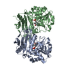 8ahrS S: Starting model for refinement |
|---|---|
| Similar structure data |
-Links
 PDBj
PDBj
- Assembly
| Deposited unit | 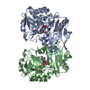
| ||||||||||||||||||
|---|---|---|---|---|---|---|---|---|---|---|---|---|---|---|---|---|---|---|---|
| 1 |
| ||||||||||||||||||
| Unit cell |
| ||||||||||||||||||
| Noncrystallographic symmetry (NCS) | NCS domain:
NCS domain segments: Component-ID: _ / Ens-ID: 1 / Beg auth comp-ID: MET / Beg label comp-ID: MET / End auth comp-ID: LEU / End label comp-ID: LEU / Refine code: _ / Auth seq-ID: 1 - 274 / Label seq-ID: 3 - 276
|
-Components
| #1: Protein | Mass: 30976.828 Da / Num. of mol.: 2 Source method: isolated from a genetically manipulated source Source: (gene. exp.) Aminobacterium colombiense (bacteria) / Strain: DSM 12261 / ALA-1 / Gene: Amico_1844 / Production host: #2: Chemical | ChemComp-LCS / [ |   Mass: 331.219 Da / Num. of mol.: 1 / Source method: obtained synthetically / Formula: C11H14N3O7P / Feature type: SUBJECT OF INVESTIGATION Mass: 331.219 Da / Num. of mol.: 1 / Source method: obtained synthetically / Formula: C11H14N3O7P / Feature type: SUBJECT OF INVESTIGATION#3: Chemical |   Mass: 248.173 Da / Num. of mol.: 2 / Source method: obtained synthetically / Formula: C8H13N2O5P / Feature type: SUBJECT OF INVESTIGATION Mass: 248.173 Da / Num. of mol.: 2 / Source method: obtained synthetically / Formula: C8H13N2O5P / Feature type: SUBJECT OF INVESTIGATION#4: Chemical | ChemComp-M7L / | 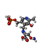  Mass: 349.234 Da / Num. of mol.: 1 / Source method: obtained synthetically / Formula: C11H16N3O8P / Feature type: SUBJECT OF INVESTIGATION Mass: 349.234 Da / Num. of mol.: 1 / Source method: obtained synthetically / Formula: C11H16N3O8P / Feature type: SUBJECT OF INVESTIGATION#5: Water | ChemComp-HOH / |  Mass: 18.015 Da / Num. of mol.: 237 / Source method: isolated from a natural source / Formula: H2O Mass: 18.015 Da / Num. of mol.: 237 / Source method: isolated from a natural source / Formula: H2OHas ligand of interest | Y | |
|---|
-Experimental details
-Experiment
| Experiment | Method: X-RAY DIFFRACTION / Number of used crystals: 1 |
|---|
- Sample preparation
| Crystal | Density Matthews: 2.26 Å3/Da / Density % sol: 45.52 % |
|---|---|
| Crystal grow | Temperature: 288 K / Method: vapor diffusion, hanging drop Details: 0.2M NaNitrate, 0.1M Bis-tris propane pH 6,5, 20% PEG3350 |
-Data collection
| Diffraction | Mean temperature: 100 K / Serial crystal experiment: N | ||||||||||||||||||||||||||||||
|---|---|---|---|---|---|---|---|---|---|---|---|---|---|---|---|---|---|---|---|---|---|---|---|---|---|---|---|---|---|---|---|
| Diffraction source | Source: ROTATING ANODE / Type: RIGAKU / Wavelength: 1.54184 Å | ||||||||||||||||||||||||||||||
| Detector | Type: RIGAKU HyPix-6000HE / Detector: PIXEL / Date: Jun 20, 2022 | ||||||||||||||||||||||||||||||
| Radiation | Protocol: SINGLE WAVELENGTH / Monochromatic (M) / Laue (L): M / Scattering type: x-ray | ||||||||||||||||||||||||||||||
| Radiation wavelength | Wavelength: 1.54184 Å / Relative weight: 1 | ||||||||||||||||||||||||||||||
| Reflection | Resolution: 1.9→21.15 Å / Num. obs: 44377 / % possible obs: 98.9 % / Redundancy: 9.5 % / CC1/2: 0.995 / Rmerge(I) obs: 0.169 / Rpim(I) all: 0.057 / Rrim(I) all: 0.178 / Net I/σ(I): 13.9 / Num. measured all: 422464 | ||||||||||||||||||||||||||||||
| Reflection shell | Diffraction-ID: 1
|
- Processing
| Software |
| ||||||||||||||||||||||||||||||||||||||||||||||||||||||||||||
|---|---|---|---|---|---|---|---|---|---|---|---|---|---|---|---|---|---|---|---|---|---|---|---|---|---|---|---|---|---|---|---|---|---|---|---|---|---|---|---|---|---|---|---|---|---|---|---|---|---|---|---|---|---|---|---|---|---|---|---|---|---|
| Refinement | Method to determine structure: MOLECULAR REPLACEMENT Starting model: 8AHR Resolution: 1.9→21.15 Å / Cor.coef. Fo:Fc: 0.955 / Cor.coef. Fo:Fc free: 0.93 / WRfactor Rfree: 0.2127 / WRfactor Rwork: 0.1739 / FOM work R set: 0.718 / SU B: 6.007 / SU ML: 0.158 / SU R Cruickshank DPI: 0.1905 / SU Rfree: 0.1713 / Cross valid method: THROUGHOUT / σ(F): 0 / ESU R: 0.191 / ESU R Free: 0.171 / Stereochemistry target values: MAXIMUM LIKELIHOOD Details: HYDROGENS HAVE BEEN ADDED IN THE RIDING POSITIONS U VALUES : REFINED INDIVIDUALLY
| ||||||||||||||||||||||||||||||||||||||||||||||||||||||||||||
| Solvent computation | Ion probe radii: 0.8 Å / Shrinkage radii: 0.8 Å / VDW probe radii: 1.2 Å / Solvent model: MASK | ||||||||||||||||||||||||||||||||||||||||||||||||||||||||||||
| Displacement parameters | Biso max: 94.24 Å2 / Biso mean: 30.653 Å2 / Biso min: 14.08 Å2
| ||||||||||||||||||||||||||||||||||||||||||||||||||||||||||||
| Refinement step | Cycle: final / Resolution: 1.9→21.15 Å
| ||||||||||||||||||||||||||||||||||||||||||||||||||||||||||||
| Refine LS restraints |
| ||||||||||||||||||||||||||||||||||||||||||||||||||||||||||||
| Refine LS restraints NCS | Ens-ID: 1 / Number: 8547 / Refine-ID: X-RAY DIFFRACTION / Type: interatomic distance / Rms dev position: 0.1 Å / Weight position: 0.05
| ||||||||||||||||||||||||||||||||||||||||||||||||||||||||||||
| LS refinement shell | Resolution: 1.9→1.949 Å / Rfactor Rfree error: 0 / Total num. of bins used: 20
|
