 Movie
Movie Controller

[English] 日本語
 Yorodumi
- PDB-8ai3: Crystal structure of radical SAM epimerase EpeE C223A mutant from... -
+ Open data

- Basic information
| Entry | Database: PDB / ID: 8ai3 | |||||||||
|---|---|---|---|---|---|---|---|---|---|---|
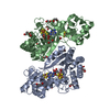
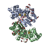
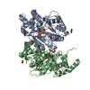
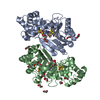
| Title | Crystal structure of radical SAM epimerase EpeE C223A mutant from Bacillus subtilis with [4Fe-4S] clusters and S-adenosyl-L-methionine bound | |||||||||
 Components Components | Putative peptide biosynthesis protein YydG | |||||||||
 Keywords Keywords | METAL BINDING PROTEIN / radical SAM / metalloenzyme / iron-sulfur / RiPP | |||||||||
| Function / homology |  Function and homology information Function and homology informationcatalytic activity / 4 iron, 4 sulfur cluster binding / metal ion binding Similarity search - Function | |||||||||
| Biological species |  | |||||||||
| Method |  X-RAY DIFFRACTION / SYNCHROTRON / MOLECULAR REPLACEMENT / Resolution: 2.1 Å X-RAY DIFFRACTION / SYNCHROTRON / MOLECULAR REPLACEMENT / Resolution: 2.1 Å | |||||||||
 Authors Authors | Kubiak, X. / Chavas, L.M.G. / Legrand, P. / Polsinelli, I. / Fyfe, C.D. / Benjdia, A. / Berteau, O. | |||||||||
| Funding support |  France, European Union, 2items France, European Union, 2items
| |||||||||
 Citation Citation | Journal: Nat.Chem.Biol. / Year: 2024 Title: Structural and mechanistic basis for RiPP epimerization by a radical SAM enzyme. Authors: Kubiak, X. / Polsinelli, I. / Chavas, L.M.G. / Fyfe, C.D. / Guillot, A. / Fradale, L. / Brewee, C. / Grimaldi, S. / Gerbaud, G. / Thureau, A. / Legrand, P. / Berteau, O. / Benjdia, A. #1: Journal: Nat Chem / Year: 2017Title: Post-translational modification of ribosomally synthesized peptides by a radical SAM epimerase in Bacillus subtilis. Authors: Benjdia, A. / Guillot, A. / Ruffie, P. / Leprince, J. / Berteau, O. | |||||||||
| History |
|
- Structure visualization
| Structure viewer | Molecule: MolmilJmol/JSmol |
|---|
- Downloads & links
-Download
| PDBx/mmCIF format | 8ai3.cif.gz | 301 KB | Display | PDBx/mmCIF format |
|---|---|---|---|---|
| PDB format | pdb8ai3.ent.gz | 242.8 KB | Display | PDB format |
| PDBx/mmJSON format | 8ai3.json.gz | Tree view | PDBx/mmJSON format | |
| Others |  Other downloads Other downloads |
-Validation report
| Arichive directory | https://data.pdbj.org/pub/pdb/validation_reports/ai/8ai3ftp://data.pdbj.org/pub/pdb/validation_reports/ai/8ai3 | HTTPS FTP |
|---|
-Related structure data

-Links
 PDBj
PDBj
- Assembly
| Deposited unit | 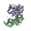
| ||||||||
|---|---|---|---|---|---|---|---|---|---|
| 1 |
| ||||||||
| Unit cell |
|
-Components
-Protein , 1 types, 2 molecules AB
| #1: Protein | Mass: 39920.379 Da / Num. of mol.: 2 / Mutation: C223A Source method: isolated from a genetically manipulated source Source: (gene. exp.) |
|---|
-Non-polymers , 5 types, 327 molecules 


| #2: Chemical | ChemComp-SF4 /  Mass: 351.640 Da / Num. of mol.: 4 / Source method: obtained synthetically / Formula: Fe4S4 / Feature type: SUBJECT OF INVESTIGATION Mass: 351.640 Da / Num. of mol.: 4 / Source method: obtained synthetically / Formula: Fe4S4 / Feature type: SUBJECT OF INVESTIGATION#3: Chemical |  Mass: 398.437 Da / Num. of mol.: 2 / Source method: obtained synthetically / Formula: C15H22N6O5S / Feature type: SUBJECT OF INVESTIGATION Mass: 398.437 Da / Num. of mol.: 2 / Source method: obtained synthetically / Formula: C15H22N6O5S / Feature type: SUBJECT OF INVESTIGATION#4: Chemical | ChemComp-EDO /  Mass: 62.068 Da / Num. of mol.: 19 / Source method: obtained synthetically / Formula: C2H6O2 Mass: 62.068 Da / Num. of mol.: 19 / Source method: obtained synthetically / Formula: C2H6O2#5: Chemical | ChemComp-GOL /  Mass: 92.094 Da / Num. of mol.: 4 / Source method: obtained synthetically / Formula: C3H8O3 Mass: 92.094 Da / Num. of mol.: 4 / Source method: obtained synthetically / Formula: C3H8O3#6: Water | ChemComp-HOH / | Mass: 18.015 Da / Num. of mol.: 298 / Source method: isolated from a natural source / Formula: H2O |
|---|
-Details
| Has ligand of interest | Y |
|---|
-Experimental details
-Experiment
| Experiment | Method: X-RAY DIFFRACTION / Number of used crystals: 1 |
|---|
- Sample preparation
| Crystal | Density Matthews: 2.98 Å3/Da / Density % sol: 58.73 % |
|---|---|
| Crystal grow | Temperature: 293 K / Method: vapor diffusion, hanging drop / pH: 7.5 Details: 12% (w/v) PEG 8000, 10% (v/v) Ethylene glycol, 0.1 M Hepes |
-Data collection
| Diffraction | Mean temperature: 100 K / Serial crystal experiment: N |
|---|---|
| Diffraction source | Source: SYNCHROTRON / Site: SOLEIL / Beamline: PROXIMA 1 / Wavelength: 0.97857 Å |
| Detector | Type: DECTRIS EIGER X 16M / Detector: PIXEL / Date: Oct 5, 2017 / Details: KB Mirrors |
| Radiation | Monochromator: channel-cut Si(111) / Protocol: SINGLE WAVELENGTH / Monochromatic (M) / Laue (L): M / Scattering type: x-ray |
| Radiation wavelength | Wavelength: 0.97857 Å / Relative weight: 1 |
| Reflection | Resolution: 2.1→46.55 Å / Num. obs: 416209 / % possible obs: 99.7 % / Redundancy: 7.4 % / CC1/2: 0.998 / Rmerge(I) obs: 0.077 / Rpim(I) all: 0.03 / Rrim(I) all: 0.082 / Net I/σ(I): 12.1 |
| Reflection shell | Resolution: 2.1→2.15 Å / Rmerge(I) obs: 1.891 / Mean I/σ(I) obs: 1 / Num. unique obs: 28724 / CC1/2: 0.509 / Rpim(I) all: 0.74 / Rrim(I) all: 2.035 |
- Processing
| Software |
| |||||||||||||||||||||||||||||||||||||||||||||||||||||||||||||||||||||||||||
|---|---|---|---|---|---|---|---|---|---|---|---|---|---|---|---|---|---|---|---|---|---|---|---|---|---|---|---|---|---|---|---|---|---|---|---|---|---|---|---|---|---|---|---|---|---|---|---|---|---|---|---|---|---|---|---|---|---|---|---|---|---|---|---|---|---|---|---|---|---|---|---|---|---|---|---|---|
| Refinement | Method to determine structure: MOLECULAR REPLACEMENT Starting model: Fe-SAD Exprimental model Resolution: 2.1→40.27 Å / Cor.coef. Fo:Fc: 0.958 / Cor.coef. Fo:Fc free: 0.958 / SU R Cruickshank DPI: 0.17 / Cross valid method: THROUGHOUT / SU R Blow DPI: 0.174 / SU Rfree Blow DPI: 0.14 / SU Rfree Cruickshank DPI: 0.139
| |||||||||||||||||||||||||||||||||||||||||||||||||||||||||||||||||||||||||||
| Displacement parameters | Biso mean: 64.84 Å2
| |||||||||||||||||||||||||||||||||||||||||||||||||||||||||||||||||||||||||||
| Refine analyze | Luzzati coordinate error obs: 0.27 Å | |||||||||||||||||||||||||||||||||||||||||||||||||||||||||||||||||||||||||||
| Refinement step | Cycle: LAST / Resolution: 2.1→40.27 Å
| |||||||||||||||||||||||||||||||||||||||||||||||||||||||||||||||||||||||||||
| Refine LS restraints |
| |||||||||||||||||||||||||||||||||||||||||||||||||||||||||||||||||||||||||||
| LS refinement shell | Resolution: 2.1→2.11 Å
| |||||||||||||||||||||||||||||||||||||||||||||||||||||||||||||||||||||||||||
| Refinement TLS params. | Refine-ID: X-RAY DIFFRACTION
| |||||||||||||||||||||||||||||||||||||||||||||||||||||||||||||||||||||||||||
| Refinement TLS group |
|
