 Movie
Movie Controller

[English] 日本語
 Yorodumi
- PDB-8ahf: PAC-FragmentDEL: Photoactivated covalent capture of DNA encoded f... -
+ Open data

- Basic information
| Entry | Database: PDB / ID: 8ahf | ||||||
|---|---|---|---|---|---|---|---|
| Title | PAC-FragmentDEL: Photoactivated covalent capture of DNA encoded fragments for hit discovery | ||||||
 Components Components | UDP-N-acetylglucosamine 2-epimerase | ||||||
 Keywords Keywords | ISOMERASE / INHIBITOR / COMPLEX / EPIMERASE / TECHNOLOGY | ||||||
| Function / homology | UDP-N-acetylglucosamine 2-epimerase (non-hydrolysing) / UDP-N-acetylglucosamine 2-epimerase WecB-like / UDP-N-acetylglucosamine 2-epimerase activity / UDP-N-acetylglucosamine 2-epimerase domain / UDP-N-acetylglucosamine 2-epimerase / nucleotide binding / Chem-M3U / UDP-N-acetylglucosamine 2-epimerase (non-hydrolyzing) Function and homology information Function and homology information | ||||||
| Biological species |  | ||||||
| Method |  X-RAY DIFFRACTION / SYNCHROTRON / MOLECULAR REPLACEMENT / Resolution: 2.271 Å X-RAY DIFFRACTION / SYNCHROTRON / MOLECULAR REPLACEMENT / Resolution: 2.271 Å | ||||||
 Authors Authors | Baker, L.M. / Murray, J.B. / Hubbard, R.E. | ||||||
| Funding support | 1items
| ||||||
 Citation Citation | Journal: Rsc Med Chem / Year: 2022 Title: PAC-FragmentDEL - photoactivated covalent capture of DNA-encoded fragments for hit discovery. Authors: Ma, H. / Murray, J.B. / Luo, H. / Cheng, X. / Chen, Q. / Song, C. / Duan, C. / Tan, P. / Zhang, L. / Liu, J. / Morgan, B.A. / Li, J. / Wan, J. / Baker, L.M. / Finnie, W. / Guetzoyan, L. / ...Authors: Ma, H. / Murray, J.B. / Luo, H. / Cheng, X. / Chen, Q. / Song, C. / Duan, C. / Tan, P. / Zhang, L. / Liu, J. / Morgan, B.A. / Li, J. / Wan, J. / Baker, L.M. / Finnie, W. / Guetzoyan, L. / Harris, R. / Hendrickson, N. / Matassova, N. / Simmonite, H. / Smith, J. / Hubbard, R.E. / Liu, G. | ||||||
| History |
|
- Structure visualization
| Structure viewer | Molecule: MolmilJmol/JSmol |
|---|
- Downloads & links
-Download
| PDBx/mmCIF format | 8ahf.cif.gz | 302.8 KB | Display | PDBx/mmCIF format |
|---|---|---|---|---|
| PDB format | pdb8ahf.ent.gz | 240.4 KB | Display | PDB format |
| PDBx/mmJSON format | 8ahf.json.gz | Tree view | PDBx/mmJSON format | |
| Others |  Other downloads Other downloads |
-Validation report
| Arichive directory | https://data.pdbj.org/pub/pdb/validation_reports/ah/8ahfftp://data.pdbj.org/pub/pdb/validation_reports/ah/8ahf | HTTPS FTP |
|---|
-Related structure data
| Related structure data | 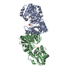 8aheC  8ahgC  8ahhC 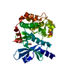 8ahiC 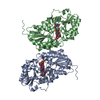 3beoS S: Starting model for refinement C: citing same article ( |
|---|---|
| Similar structure data |
-Links
 PDBj
PDBj- Assembly
| Deposited unit | 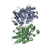
| ||||||||||||
|---|---|---|---|---|---|---|---|---|---|---|---|---|---|
| 1 | 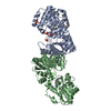
| ||||||||||||
| Unit cell |
| ||||||||||||
| Components on special symmetry positions |
|
-Components
| #1: Protein | Mass: 42062.078 Da / Num. of mol.: 2 Source method: isolated from a genetically manipulated source Source: (gene. exp.) References: UniProt: A0A348ACF3, UDP-N-acetylglucosamine 2-epimerase (non-hydrolysing) #2: Chemical |   Mass: 339.297 Da / Num. of mol.: 2 / Source method: obtained synthetically / Formula: C14H15F2N5O3 / Feature type: SUBJECT OF INVESTIGATION Mass: 339.297 Da / Num. of mol.: 2 / Source method: obtained synthetically / Formula: C14H15F2N5O3 / Feature type: SUBJECT OF INVESTIGATION#3: Chemical | ChemComp-SO4 /   Mass: 96.063 Da / Num. of mol.: 8 / Source method: obtained synthetically / Formula: SO4 Mass: 96.063 Da / Num. of mol.: 8 / Source method: obtained synthetically / Formula: SO4#4: Water | ChemComp-HOH / |  Mass: 18.015 Da / Num. of mol.: 248 / Source method: isolated from a natural source / Formula: H2O Mass: 18.015 Da / Num. of mol.: 248 / Source method: isolated from a natural source / Formula: H2OHas ligand of interest | Y | |
|---|
-Experimental details
-Experiment
| Experiment | Method: X-RAY DIFFRACTION / Number of used crystals: 1 |
|---|
- Sample preparation
| Crystal | Density Matthews: 2.37 Å3/Da / Density % sol: 47.6 % / Description: Needles |
|---|---|
| Crystal grow | Temperature: 292 K / Method: vapor diffusion, hanging drop / pH: 5.6 Details: 0.2 M Ammonium sulfate 50 mM BIS-TRIS pH 5.6 28% PEG 3350 |
-Data collection
| Diffraction | Mean temperature: 100 K / Serial crystal experiment: N |
|---|---|
| Diffraction source | Source: SYNCHROTRON / Site: Diamond  / Beamline: I03 / Wavelength: 0.9763 Å / Beamline: I03 / Wavelength: 0.9763 Å |
| Detector | Type: DECTRIS EIGER2 XE 16M / Detector: PIXEL / Date: Nov 27, 2021 |
| Radiation | Monochromator: Laue / Protocol: SINGLE WAVELENGTH / Monochromatic (M) / Laue (L): M / Scattering type: x-ray |
| Radiation wavelength | Wavelength: 0.9763 Å / Relative weight: 1 |
| Reflection | Resolution: 2.271→85.17 Å / Num. obs: 36587 / % possible obs: 95.7 % / Redundancy: 26.8 % / CC1/2: 0.999 / Rmerge(I) obs: 0.201 / Net I/σ(I): 14.6 |
| Reflection shell | Resolution: 2.271→2.353 Å / Redundancy: 27.7 % / Rmerge(I) obs: 3.536 / Mean I/σ(I) obs: 1.2 / Num. unique obs: 1828 / CC1/2: 0.59 / % possible all: 52.8 |
- Processing
| Software |
| |||||||||||||||||||||||||||||||||||||||||||||||||||||||||||||||||||||||||||||||||||||||||||||||||||||||||||||||||||||||||||||||||||||||||||||||||||||||||||||||||||||||||||||||||||||||||||||||||||||||||||||||||||||||||||||||||||||||
|---|---|---|---|---|---|---|---|---|---|---|---|---|---|---|---|---|---|---|---|---|---|---|---|---|---|---|---|---|---|---|---|---|---|---|---|---|---|---|---|---|---|---|---|---|---|---|---|---|---|---|---|---|---|---|---|---|---|---|---|---|---|---|---|---|---|---|---|---|---|---|---|---|---|---|---|---|---|---|---|---|---|---|---|---|---|---|---|---|---|---|---|---|---|---|---|---|---|---|---|---|---|---|---|---|---|---|---|---|---|---|---|---|---|---|---|---|---|---|---|---|---|---|---|---|---|---|---|---|---|---|---|---|---|---|---|---|---|---|---|---|---|---|---|---|---|---|---|---|---|---|---|---|---|---|---|---|---|---|---|---|---|---|---|---|---|---|---|---|---|---|---|---|---|---|---|---|---|---|---|---|---|---|---|---|---|---|---|---|---|---|---|---|---|---|---|---|---|---|---|---|---|---|---|---|---|---|---|---|---|---|---|---|---|---|---|---|---|---|---|---|---|---|---|---|---|---|---|---|---|---|---|---|
| Refinement | Method to determine structure: MOLECULAR REPLACEMENT Starting model: 3BEO Resolution: 2.271→85.17 Å / Cor.coef. Fo:Fc: 0.96 / Cor.coef. Fo:Fc free: 0.928 / SU B: 7.088 / SU ML: 0.169 / Cross valid method: FREE R-VALUE / ESU R: 0.322 / ESU R Free: 0.235 Details: Hydrogens have been added in their riding positions
| |||||||||||||||||||||||||||||||||||||||||||||||||||||||||||||||||||||||||||||||||||||||||||||||||||||||||||||||||||||||||||||||||||||||||||||||||||||||||||||||||||||||||||||||||||||||||||||||||||||||||||||||||||||||||||||||||||||||
| Solvent computation | Ion probe radii: 0.8 Å / Shrinkage radii: 0.8 Å / VDW probe radii: 1.2 Å / Solvent model: MASK BULK SOLVENT | |||||||||||||||||||||||||||||||||||||||||||||||||||||||||||||||||||||||||||||||||||||||||||||||||||||||||||||||||||||||||||||||||||||||||||||||||||||||||||||||||||||||||||||||||||||||||||||||||||||||||||||||||||||||||||||||||||||||
| Displacement parameters | Biso mean: 48.125 Å2
| |||||||||||||||||||||||||||||||||||||||||||||||||||||||||||||||||||||||||||||||||||||||||||||||||||||||||||||||||||||||||||||||||||||||||||||||||||||||||||||||||||||||||||||||||||||||||||||||||||||||||||||||||||||||||||||||||||||||
| Refinement step | Cycle: LAST / Resolution: 2.271→85.17 Å
| |||||||||||||||||||||||||||||||||||||||||||||||||||||||||||||||||||||||||||||||||||||||||||||||||||||||||||||||||||||||||||||||||||||||||||||||||||||||||||||||||||||||||||||||||||||||||||||||||||||||||||||||||||||||||||||||||||||||
| Refine LS restraints |
| |||||||||||||||||||||||||||||||||||||||||||||||||||||||||||||||||||||||||||||||||||||||||||||||||||||||||||||||||||||||||||||||||||||||||||||||||||||||||||||||||||||||||||||||||||||||||||||||||||||||||||||||||||||||||||||||||||||||
| LS refinement shell | Refine-ID: X-RAY DIFFRACTION / Total num. of bins used: 20
|
