+ Open data
- Basic information
| Entry | Database: PDB / ID: 8afw | ||||||
|---|---|---|---|---|---|---|---|
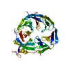
| Title | Tube assembly of Atg18-WT | ||||||
 Components Components | Autophagy-related protein 18 | ||||||
 Keywords Keywords | MEMBRANE PROTEIN / autophagy / membrane remodeling / PIP binding / PI3P / PI(3 / 5)P2 / lipid binding protein | ||||||
| Function / homology |  Function and homology information Function and homology informationregulation of phosphatidylinositol biosynthetic process / PAS complex / 1-phosphatidyl-1D-myo-inositol 3,5-bisphosphate metabolic process / phagophore / positive regulation of vacuole organization / vacuolar protein processing / Macroautophagy / glycophagy / cytoplasm to vacuole targeting by the Cvt pathway / nucleophagy ...regulation of phosphatidylinositol biosynthetic process / PAS complex / 1-phosphatidyl-1D-myo-inositol 3,5-bisphosphate metabolic process / phagophore / positive regulation of vacuole organization / vacuolar protein processing / Macroautophagy / glycophagy / cytoplasm to vacuole targeting by the Cvt pathway / nucleophagy / protein localization to phagophore assembly site / phagophore assembly site membrane / late endosome to vacuole transport / autophagy of mitochondrion / pexophagy / piecemeal microautophagy of the nucleus / phosphatidylinositol-3-phosphate binding / fungal-type vacuole membrane / phagophore assembly site / phosphatidylinositol-4-phosphate binding / phosphatidylinositol-3,5-bisphosphate binding / vacuolar membrane / autophagosome assembly / ubiquitin binding / cell periphery / macroautophagy / protein-macromolecule adaptor activity / endosome membrane / endosome / protein-containing complex / cytosol Similarity search - Function | ||||||
| Biological species |  | ||||||
| Method | ELECTRON MICROSCOPY / helical reconstruction / cryo EM / Resolution: 3.8 Å | ||||||
 Authors Authors | Mann, D. / Fromm, S. / Martinez-Sanchez, A. / Gopaldass, N. / Mayer, A. / Sachse, C. | ||||||
| Funding support |  Germany, 1items Germany, 1items
| ||||||
 Citation Citation |  Journal: Nat Commun / Year: 2023 Journal: Nat Commun / Year: 2023Title: Atg18 oligomer organization in assembled tubes and on lipid membrane scaffolds. Authors: Daniel Mann / Simon A Fromm / Antonio Martinez-Sanchez / Navin Gopaldass / Ramona Choy / Andreas Mayer / Carsten Sachse /    Abstract: Autophagy-related protein 18 (Atg18) participates in the elongation of early autophagosomal structures in concert with Atg2 and Atg9 complexes. How Atg18 contributes to the structural coordination of ...Autophagy-related protein 18 (Atg18) participates in the elongation of early autophagosomal structures in concert with Atg2 and Atg9 complexes. How Atg18 contributes to the structural coordination of Atg2 and Atg9 at the isolation membrane remains to be understood. Here, we determined the cryo-EM structures of Atg18 organized in helical tubes, Atg18 oligomers in solution as well as on lipid membrane scaffolds. The helical assembly is composed of Atg18 tetramers forming a lozenge cylindrical lattice with remarkable structural similarity to the COPII outer coat. When reconstituted with lipid membranes, using subtomogram averaging we determined tilted Atg18 dimer structures bridging two juxtaposed lipid membranes spaced apart by 80 Å. Moreover, lipid reconstitution experiments further delineate the contributions of Atg18's FRRG motif and the amphipathic helical extension in membrane interaction. The observed structural plasticity of Atg18's oligomeric organization and membrane binding properties provide a molecular framework for the positioning of downstream components of the autophagy machinery. | ||||||
| History |
|
- Structure visualization
| Structure viewer | Molecule:  Molmil Molmil Jmol/JSmol Jmol/JSmol |
|---|
- Downloads & links
- Download
| PDBx/mmCIF format |  8afw.cif.gz 8afw.cif.gz | 311.1 KB | Display |  PDBx/mmCIF format PDBx/mmCIF format |
|---|---|---|---|---|
| PDB format |  pdb8afw.ent.gz pdb8afw.ent.gz | 236.1 KB | Display |  PDB format PDB format |
| PDBx/mmJSON format |  8afw.json.gz 8afw.json.gz | Tree view |  PDBx/mmJSON format PDBx/mmJSON format | |
| Others |  Other downloads Other downloads |
-Validation report
| Summary document |  8afw_validation.pdf.gz 8afw_validation.pdf.gz | 1.5 MB | Display |  wwPDB validaton report wwPDB validaton report |
|---|---|---|---|---|
| Full document |  8afw_full_validation.pdf.gz 8afw_full_validation.pdf.gz | 1.5 MB | Display | |
| Data in XML |  8afw_validation.xml.gz 8afw_validation.xml.gz | 55.1 KB | Display | |
| Data in CIF |  8afw_validation.cif.gz 8afw_validation.cif.gz | 79.9 KB | Display | |
| Arichive directory |  https://data.pdbj.org/pub/pdb/validation_reports/af/8afw https://data.pdbj.org/pub/pdb/validation_reports/af/8afw ftp://data.pdbj.org/pub/pdb/validation_reports/af/8afw ftp://data.pdbj.org/pub/pdb/validation_reports/af/8afw | HTTPS FTP |
-Related structure data
| Related structure data |  15410MC  8afqC  8afxC  8afyC M: map data used to model this data C: citing same article ( |
|---|---|
| Similar structure data | Similarity search - Function & homology  F&H Search F&H Search |
- Links
Links
- Assembly
| Deposited unit | 
|
|---|---|
| 1 | x 20
|
- Components
| #1: Protein | Mass: 55157.930 Da / Num. of mol.: 4 Source method: isolated from a genetically manipulated source Source: (gene. exp.)  Strain: ATCC 204508 / S288c / Gene: ATG18, AUT10, CVT18, NMR1, SVP1, YFR021W / Production host:  |
|---|
-Experimental details
-Experiment
| Experiment | Method: ELECTRON MICROSCOPY |
|---|---|
| EM experiment | Aggregation state: FILAMENT / 3D reconstruction method: helical reconstruction |
- Sample preparation
| Component | Name: Filament assembly of Atg18-WT / Type: COMPLEX / Entity ID: all / Source: RECOMBINANT |
|---|---|
| Molecular weight | Experimental value: NO |
| Source (natural) | Organism:  |
| Source (recombinant) | Organism:  |
| Buffer solution | pH: 7.2 |
| Specimen | Embedding applied: NO / Shadowing applied: NO / Staining applied: NO / Vitrification applied: YES |
| Vitrification | Instrument: LEICA EM GP / Cryogen name: ETHANE / Humidity: 99 % / Chamber temperature: 291 K |
- Electron microscopy imaging
| Experimental equipment |  Model: Titan Krios / Image courtesy: FEI Company |
|---|---|
| Microscopy | Model: TFS KRIOS |
| Electron gun | Electron source:  FIELD EMISSION GUN / Accelerating voltage: 300 kV / Illumination mode: FLOOD BEAM FIELD EMISSION GUN / Accelerating voltage: 300 kV / Illumination mode: FLOOD BEAM |
| Electron lens | Mode: BRIGHT FIELD / Nominal defocus max: 3000 nm / Nominal defocus min: 1000 nm / Alignment procedure: COMA FREE |
| Specimen holder | Cryogen: NITROGEN / Specimen holder model: FEI TITAN KRIOS AUTOGRID HOLDER |
| Image recording | Electron dose: 90 e/Å2 / Detector mode: COUNTING / Film or detector model: GATAN K2 SUMMIT (4k x 4k) / Num. of real images: 1812 |
- Processing
| EM software |
| |||||||||||||||||||||||||||
|---|---|---|---|---|---|---|---|---|---|---|---|---|---|---|---|---|---|---|---|---|---|---|---|---|---|---|---|---|
| CTF correction | Type: PHASE FLIPPING AND AMPLITUDE CORRECTION | |||||||||||||||||||||||||||
| Helical symmerty | Angular rotation/subunit: 70 ° / Axial rise/subunit: 19.4 Å / Axial symmetry: C1 | |||||||||||||||||||||||||||
| 3D reconstruction | Resolution: 3.8 Å / Resolution method: FSC 0.143 CUT-OFF / Num. of particles: 133981 / Symmetry type: HELICAL | |||||||||||||||||||||||||||
| Atomic model building | Protocol: RIGID BODY FIT / Space: REAL / Target criteria: Correlation coefficient Details: Initial PDB was derived from the Atg18-PR72AA filament structure, subunits were individually docked inside the density and the PR72AA loop was manually modified in Coot |
 Movie
Movie Controller


 PDBj
PDBj
