 Movie
Movie Controller

[English] 日本語
 Yorodumi
- PDB-8afv: DaArgC3 - Engineered Formyl Phosphate Reductase with 3 substituti... -
+ Open data

- Basic information
| Entry | Database: PDB / ID: 8afv | |||||||||
|---|---|---|---|---|---|---|---|---|---|---|
| Title | DaArgC3 - Engineered Formyl Phosphate Reductase with 3 substitutions (S178V, G182V, L233I) | |||||||||
 Components Components | N-acetyl-gamma-glutamyl-phosphate reductase | |||||||||
 Keywords Keywords | OXIDOREDUCTASE / formyl phosphate | |||||||||
| Function / homology |  Function and homology information Function and homology informationN-acetyl-gamma-glutamyl-phosphate reductase / N-acetyl-gamma-glutamyl-phosphate reductase activity / L-arginine biosynthetic process / NADP+ binding / NAD binding / cytoplasm Similarity search - Function | |||||||||
| Biological species |  Denitrovibrio acetiphilus DSM 12809 (bacteria) Denitrovibrio acetiphilus DSM 12809 (bacteria) | |||||||||
| Method |  X-RAY DIFFRACTION / SYNCHROTRON / MOLECULAR REPLACEMENT / Resolution: 2.19 Å X-RAY DIFFRACTION / SYNCHROTRON / MOLECULAR REPLACEMENT / Resolution: 2.19 Å | |||||||||
 Authors Authors | Pfister, P. / Nattermann, M. / Zarzycki, J. / Erb, T.J. | |||||||||
| Funding support |  Germany, 2items Germany, 2items
| |||||||||
 Citation Citation | Journal: Nat Commun / Year: 2023 Title: Engineering a new-to-nature cascade for phosphate-dependent formate to formaldehyde conversion in vitro and in vivo. Authors: Nattermann, M. / Wenk, S. / Pfister, P. / He, H. / Lee, S.H. / Szymanski, W. / Guntermann, N. / Zhu, F. / Nickel, L. / Wallner, C. / Zarzycki, J. / Paczia, N. / Gaissert, N. / Francio, G. / ...Authors: Nattermann, M. / Wenk, S. / Pfister, P. / He, H. / Lee, S.H. / Szymanski, W. / Guntermann, N. / Zhu, F. / Nickel, L. / Wallner, C. / Zarzycki, J. / Paczia, N. / Gaissert, N. / Francio, G. / Leitner, W. / Gonzalez, R. / Erb, T.J. | |||||||||
| History |
|
- Structure visualization
| Structure viewer | Molecule: MolmilJmol/JSmol |
|---|
- Downloads & links
-Download
| PDBx/mmCIF format | 8afv.cif.gz | 521.9 KB | Display | PDBx/mmCIF format |
|---|---|---|---|---|
| PDB format | pdb8afv.ent.gz | 431 KB | Display | PDB format |
| PDBx/mmJSON format | 8afv.json.gz | Tree view | PDBx/mmJSON format | |
| Others |  Other downloads Other downloads |
-Validation report
| Arichive directory | https://data.pdbj.org/pub/pdb/validation_reports/af/8afvftp://data.pdbj.org/pub/pdb/validation_reports/af/8afv | HTTPS FTP |
|---|
-Related structure data
| Related structure data |  8afuC 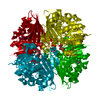 3dr3S S: Starting model for refinement C: citing same article ( |
|---|---|
| Similar structure data |
-Links
 PDBj
PDBj- Assembly
| Deposited unit | 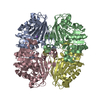
| ||||||||
|---|---|---|---|---|---|---|---|---|---|
| 1 | 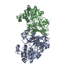
| ||||||||
| 2 | 
| ||||||||
| Unit cell |
|
-Components
| #1: Protein | Mass: 38001.469 Da / Num. of mol.: 4 Source method: isolated from a genetically manipulated source Source: (gene. exp.) Denitrovibrio acetiphilus DSM 12809 (bacteria)Strain: DSM 12809 / NBRC 114555 / N2460 / Gene: argC, Dacet_0460 / Production host: References: UniProt: D4H3H4, N-acetyl-gamma-glutamyl-phosphate reductase #2: Chemical | ChemComp-NA /   Mass: 22.990 Da / Num. of mol.: 4 / Source method: obtained synthetically / Formula: Na Mass: 22.990 Da / Num. of mol.: 4 / Source method: obtained synthetically / Formula: Na#3: Water | ChemComp-HOH / |  Mass: 18.015 Da / Num. of mol.: 534 / Source method: isolated from a natural source / Formula: H2O Mass: 18.015 Da / Num. of mol.: 534 / Source method: isolated from a natural source / Formula: H2OHas ligand of interest | N | Has protein modification | N | |
|---|
-Experimental details
-Experiment
| Experiment | Method: X-RAY DIFFRACTION / Number of used crystals: 1 |
|---|
- Sample preparation
| Crystal | Density Matthews: 2.33 Å3/Da / Density % sol: 47.23 % |
|---|---|
| Crystal grow | Temperature: 288.15 K / Method: vapor diffusion, sitting drop / pH: 7.5 Details: 0.2 M Sodium chloride 0.1 M HEPES pH 7.5 25%(w/v) PEG 4000 |
-Data collection
| Diffraction | Mean temperature: 80 K / Ambient temp details: cryostream / Serial crystal experiment: N | |||||||||||||||||||||||||||||||||||||||||||||||||||||||||||||||||||||||||||||||||||||||||||||||||||
|---|---|---|---|---|---|---|---|---|---|---|---|---|---|---|---|---|---|---|---|---|---|---|---|---|---|---|---|---|---|---|---|---|---|---|---|---|---|---|---|---|---|---|---|---|---|---|---|---|---|---|---|---|---|---|---|---|---|---|---|---|---|---|---|---|---|---|---|---|---|---|---|---|---|---|---|---|---|---|---|---|---|---|---|---|---|---|---|---|---|---|---|---|---|---|---|---|---|---|---|---|
| Diffraction source | Source: SYNCHROTRON / Site: ESRF  / Beamline: ID30B / Wavelength: 0.97625 Å / Beamline: ID30B / Wavelength: 0.97625 Å | |||||||||||||||||||||||||||||||||||||||||||||||||||||||||||||||||||||||||||||||||||||||||||||||||||
| Detector | Type: DECTRIS PILATUS 6M / Detector: PIXEL / Date: Feb 26, 2022 | |||||||||||||||||||||||||||||||||||||||||||||||||||||||||||||||||||||||||||||||||||||||||||||||||||
| Radiation | Protocol: SINGLE WAVELENGTH / Monochromatic (M) / Laue (L): M / Scattering type: x-ray | |||||||||||||||||||||||||||||||||||||||||||||||||||||||||||||||||||||||||||||||||||||||||||||||||||
| Radiation wavelength | Wavelength: 0.97625 Å / Relative weight: 1 | |||||||||||||||||||||||||||||||||||||||||||||||||||||||||||||||||||||||||||||||||||||||||||||||||||
| Reflection | Resolution: 2.19→42.364 Å / Num. obs: 66917 / % possible obs: 97 % / Redundancy: 3.2 % / Biso Wilson estimate: 29.75 Å2 / Rpim(I) all: 0.035 / Rrim(I) all: 0.068 / Rsym value: 0.057 / Net I/av σ(I): 8.8 / Net I/σ(I): 13.5 | |||||||||||||||||||||||||||||||||||||||||||||||||||||||||||||||||||||||||||||||||||||||||||||||||||
| Reflection shell | Diffraction-ID: 1
|
- Processing
| Software |
| |||||||||||||||||||||||||||||||||||||||||||||||||||||||||||||||||||||||||||||||||||||||||||||||||||||||||
|---|---|---|---|---|---|---|---|---|---|---|---|---|---|---|---|---|---|---|---|---|---|---|---|---|---|---|---|---|---|---|---|---|---|---|---|---|---|---|---|---|---|---|---|---|---|---|---|---|---|---|---|---|---|---|---|---|---|---|---|---|---|---|---|---|---|---|---|---|---|---|---|---|---|---|---|---|---|---|---|---|---|---|---|---|---|---|---|---|---|---|---|---|---|---|---|---|---|---|---|---|---|---|---|---|---|---|
| Refinement | Method to determine structure: MOLECULAR REPLACEMENT Starting model: 3dr3 Resolution: 2.19→42.36 Å / SU ML: 0.19 / Cross valid method: THROUGHOUT / σ(F): 1.34 / Phase error: 19.49 / Stereochemistry target values: ML
| |||||||||||||||||||||||||||||||||||||||||||||||||||||||||||||||||||||||||||||||||||||||||||||||||||||||||
| Solvent computation | Shrinkage radii: 0.9 Å / VDW probe radii: 1.1 Å / Solvent model: FLAT BULK SOLVENT MODEL | |||||||||||||||||||||||||||||||||||||||||||||||||||||||||||||||||||||||||||||||||||||||||||||||||||||||||
| Displacement parameters | Biso max: 134.56 Å2 / Biso mean: 38.5589 Å2 / Biso min: 14.74 Å2 | |||||||||||||||||||||||||||||||||||||||||||||||||||||||||||||||||||||||||||||||||||||||||||||||||||||||||
| Refinement step | Cycle: final / Resolution: 2.19→42.36 Å
| |||||||||||||||||||||||||||||||||||||||||||||||||||||||||||||||||||||||||||||||||||||||||||||||||||||||||
| LS refinement shell | Refine-ID: X-RAY DIFFRACTION / Rfactor Rfree error: 0 / Total num. of bins used: 14
| |||||||||||||||||||||||||||||||||||||||||||||||||||||||||||||||||||||||||||||||||||||||||||||||||||||||||
| Refinement TLS params. | Method: refined / Origin x: -38.4898 Å / Origin y: -1.2472 Å / Origin z: -0.0408 Å
| |||||||||||||||||||||||||||||||||||||||||||||||||||||||||||||||||||||||||||||||||||||||||||||||||||||||||
| Refinement TLS group |
|
