 Movie
Movie Controller


+ Open data

- Basic information
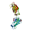
| Entry | Database: PDB / ID: 8afo | ||||||
|---|---|---|---|---|---|---|---|
| Title | Structure of fibronectin 2 and 3 of L1CAM at 2.0 Angstrom | ||||||
 Components Components | Neural cell adhesion molecule L1 | ||||||
 Keywords Keywords | CELL ADHESION / fibronectin / glycans / L1CAM | ||||||
| Function / homology |  Function and homology information Function and homology informationL1CAM interactions / axon guidance receptor activity / Interaction between L1 and Ankyrins / Basigin interactions / axon development / homophilic cell-cell adhesion / Recycling pathway of L1 / positive regulation of axon extension / axonal growth cone / axon guidance ...L1CAM interactions / axon guidance receptor activity / Interaction between L1 and Ankyrins / Basigin interactions / axon development / homophilic cell-cell adhesion / Recycling pathway of L1 / positive regulation of axon extension / axonal growth cone / axon guidance / cell-matrix adhesion / Signal transduction by L1 / synapse organization / neuron projection development / chemotaxis / cell migration / nervous system development / : / cell adhesion / protein domain specific binding / axon / focal adhesion / neuronal cell body / dendrite / cell surface / plasma membrane Similarity search - Function | ||||||
| Biological species |  Homo sapiens (human) Homo sapiens (human) | ||||||
| Method |  X-RAY DIFFRACTION / SYNCHROTRON / MOLECULAR REPLACEMENT / Resolution: 1.99 Å X-RAY DIFFRACTION / SYNCHROTRON / MOLECULAR REPLACEMENT / Resolution: 1.99 Å | ||||||
 Authors Authors | Guedez, G. / Loew, C. | ||||||
| Funding support | 1items
| ||||||
 Citation Citation | Journal: Faseb J. / Year: 2023 Title: X-ray structure and function of fibronectin domains two and three of the neural cell adhesion molecule L1. Authors: Guedez, G. / Loers, G. / Jeffries, C.M. / Kozak, S. / Meijers, R. / Svergun, D.I. / Schachner, M. / Low, C. | ||||||
| History |
|
- Structure visualization
| Structure viewer | Molecule: MolmilJmol/JSmol |
|---|
- Downloads & links
-Download
| PDBx/mmCIF format | 8afo.cif.gz | 125.3 KB | Display | PDBx/mmCIF format |
|---|---|---|---|---|
| PDB format | pdb8afo.ent.gz | 80.7 KB | Display | PDB format |
| PDBx/mmJSON format | 8afo.json.gz | Tree view | PDBx/mmJSON format | |
| Others |  Other downloads Other downloads |
-Validation report
| Arichive directory | https://data.pdbj.org/pub/pdb/validation_reports/af/8afoftp://data.pdbj.org/pub/pdb/validation_reports/af/8afo | HTTPS FTP |
|---|
-Related structure data
-Links
 PDBj
PDBj


- Assembly
| Deposited unit | 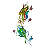
| ||||||||||||
|---|---|---|---|---|---|---|---|---|---|---|---|---|---|
| 1 |
| ||||||||||||
| Unit cell |
|
-Components
| #1: Protein | Mass: 24377.211 Da / Num. of mol.: 1 Source method: isolated from a genetically manipulated source Source: (gene. exp.) Homo sapiens (human) / Gene: L1CAM, CAML1, MIC5 / Cell line (production host): HEK293 GnTI- / Production host: Homo sapiens (human) / References: UniProt: P32004 | ||||||
|---|---|---|---|---|---|---|---|
| #2: Polysaccharide | 2-acetamido-2-deoxy-beta-D-glucopyranose-(1-4)-2-acetamido-2-deoxy-beta-D-glucopyranose Source method: isolated from a genetically manipulated source | ||||||
| #3: Sugar | ChemComp-NAG /   Type: D-saccharide, beta linking / Mass: 221.208 Da / Num. of mol.: 4 / Source method: obtained synthetically / Formula: C8H15NO6 / Feature type: SUBJECT OF INVESTIGATION Type: D-saccharide, beta linking / Mass: 221.208 Da / Num. of mol.: 4 / Source method: obtained synthetically / Formula: C8H15NO6 / Feature type: SUBJECT OF INVESTIGATION#4: Water | ChemComp-HOH / |  Mass: 18.015 Da / Num. of mol.: 168 / Source method: isolated from a natural source / Formula: H2O Mass: 18.015 Da / Num. of mol.: 168 / Source method: isolated from a natural source / Formula: H2OHas ligand of interest | Y | Has protein modification | Y | |
-Experimental details
-Experiment
| Experiment | Method: X-RAY DIFFRACTION / Number of used crystals: 1 |
|---|
- Sample preparation
| Crystal | Density Matthews: 2.95 Å3/Da / Density % sol: 58.24 % |
|---|---|
| Crystal grow | Temperature: 292 K / Method: vapor diffusion, sitting drop Details: 150 mM sodium citrate pH 5.5, 10.6% PEG 20.000, 10 mM sodium potassium tartrate |
-Data collection
| Diffraction | Mean temperature: 100 K / Serial crystal experiment: N |
|---|---|
| Diffraction source | Source: SYNCHROTRON / Site: PETRA III, EMBL c/o DESY  / Beamline: P14 (MX2) / Wavelength: 0.9762 Å / Beamline: P14 (MX2) / Wavelength: 0.9762 Å |
| Detector | Type: DECTRIS EIGER X 16M / Detector: PIXEL / Date: Nov 11, 2020 |
| Radiation | Protocol: SINGLE WAVELENGTH / Monochromatic (M) / Laue (L): M / Scattering type: x-ray |
| Radiation wavelength | Wavelength: 0.9762 Å / Relative weight: 1 |
| Reflection | Resolution: 1.99→50 Å / Num. obs: 18884 / % possible obs: 94.02 % / Redundancy: 12.4 % / Biso Wilson estimate: 43.43 Å2 / CC1/2: 0.99 / Rrim(I) all: 0.08 / Net I/σ(I): 17.8 |
| Reflection shell | Resolution: 1.99→2.1 Å / Num. unique obs: 1022 / CC1/2: 0.69 / Rrim(I) all: 1 |
- Processing
| Software |
| ||||||||||||||||||||||||||||||||||||||||||||||||||||||||
|---|---|---|---|---|---|---|---|---|---|---|---|---|---|---|---|---|---|---|---|---|---|---|---|---|---|---|---|---|---|---|---|---|---|---|---|---|---|---|---|---|---|---|---|---|---|---|---|---|---|---|---|---|---|---|---|---|---|
| Refinement | Method to determine structure: MOLECULAR REPLACEMENT / Resolution: 1.99→48.52 Å / SU ML: 0.185 / Cross valid method: FREE R-VALUE / σ(F): 1.35 / Phase error: 28.4086 Stereochemistry target values: GeoStd + Monomer Library + CDL v1.2
| ||||||||||||||||||||||||||||||||||||||||||||||||||||||||
| Solvent computation | Shrinkage radii: 0.9 Å / VDW probe radii: 1.11 Å / Solvent model: FLAT BULK SOLVENT MODEL | ||||||||||||||||||||||||||||||||||||||||||||||||||||||||
| Displacement parameters | Biso mean: 53.62 Å2 | ||||||||||||||||||||||||||||||||||||||||||||||||||||||||
| Refinement step | Cycle: LAST / Resolution: 1.99→48.52 Å
| ||||||||||||||||||||||||||||||||||||||||||||||||||||||||
| Refine LS restraints |
| ||||||||||||||||||||||||||||||||||||||||||||||||||||||||
| LS refinement shell |
| ||||||||||||||||||||||||||||||||||||||||||||||||||||||||
| Refinement TLS params. | Method: refined / Origin x: -17.7962692944 Å / Origin y: -24.4805560412 Å / Origin z: 10.7070149513 Å
| ||||||||||||||||||||||||||||||||||||||||||||||||||||||||
| Refinement TLS group | Selection details: all |
