 Movie
Movie Controller


+ Open data

- Basic information
| Entry | Database: PDB / ID: 8af1 | ||||||
|---|---|---|---|---|---|---|---|
| Title | Beta-Lytic Protease from Lysobacter capsici | ||||||
 Components Components | Peptidase M23 | ||||||
 Keywords Keywords | HYDROLASE / ELASTASE / PEPTIDOGLYCAN / Beta-lytic protease / antimicrobial activity / Lysobacter / Metalloprotease | ||||||
| Function / homology | Peptidase M23A, B-lytic metalloendopeptidase / Duplicated hybrid motif / metalloendopeptidase activity / proteolysis / FORMIC ACID / Peptidase M23 Function and homology information Function and homology information | ||||||
| Biological species |  Lysobacter capsici (bacteria) Lysobacter capsici (bacteria) | ||||||
| Method |  X-RAY DIFFRACTION / SYNCHROTRON / MOLECULAR REPLACEMENT / Resolution: 1.57 Å X-RAY DIFFRACTION / SYNCHROTRON / MOLECULAR REPLACEMENT / Resolution: 1.57 Å | ||||||
 Authors Authors | Gabdulkhakov, A.G. / Tishchenko, T.V. / Kudryakova, I.V. / Afoshin, A.S. / Vasilyeva, N.V. | ||||||
| Funding support |  Russian Federation, 1items Russian Federation, 1items
| ||||||
 Citation Citation | Journal: Int J Mol Sci / Year: 2022 Title: Structural and Functional Characterization of beta-lytic Protease from Lysobacter capsici VKM B-2533 T. Authors: Afoshin, A. / Tishchenko, S. / Gabdulkhakov, A. / Kudryakova, I. / Galemina, I. / Zelenov, D. / Leontyevskaya, E. / Saharova, S. / Leontyevskaya Vasilyeva, N. | ||||||
| History |
|
- Structure visualization
| Structure viewer | Molecule: MolmilJmol/JSmol |
|---|
- Downloads & links
-Download
| PDBx/mmCIF format | 8af1.cif.gz | 104.5 KB | Display | PDBx/mmCIF format |
|---|---|---|---|---|
| PDB format | pdb8af1.ent.gz | 64.8 KB | Display | PDB format |
| PDBx/mmJSON format | 8af1.json.gz | Tree view | PDBx/mmJSON format | |
| Others |  Other downloads Other downloads |
-Validation report
| Arichive directory | https://data.pdbj.org/pub/pdb/validation_reports/af/8af1ftp://data.pdbj.org/pub/pdb/validation_reports/af/8af1 | HTTPS FTP |
|---|
-Related structure data
| Related structure data |  3it5S S: Starting model for refinement |
|---|---|
| Similar structure data |
-Links
 PDBj
PDBj- Assembly
| Deposited unit | 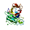
| ||||||||||||
|---|---|---|---|---|---|---|---|---|---|---|---|---|---|
| 1 |
| ||||||||||||
| Unit cell |
| ||||||||||||
| Components on special symmetry positions |
|
-Components
-Protein , 1 types, 1 molecules A
| #1: Protein | Mass: 19221.842 Da / Num. of mol.: 1 Source method: isolated from a genetically manipulated source Details: VKM B-2533T / Source: (gene. exp.) Lysobacter capsici (bacteria) / Gene: CNO08_04090 / Production host: Lysobacter capsici (bacteria) / References: UniProt: A0A290ZX82 |
|---|
-Non-polymers , 5 types, 154 molecules 


| #2: Chemical | ChemComp-ZN /  Mass: 65.409 Da / Num. of mol.: 1 / Source method: obtained synthetically / Formula: Zn / Feature type: SUBJECT OF INVESTIGATION Mass: 65.409 Da / Num. of mol.: 1 / Source method: obtained synthetically / Formula: Zn / Feature type: SUBJECT OF INVESTIGATION | ||||
|---|---|---|---|---|---|
| #3: Chemical | ChemComp-CL /  Mass: 35.453 Da / Num. of mol.: 1 / Source method: obtained synthetically / Formula: Cl Mass: 35.453 Da / Num. of mol.: 1 / Source method: obtained synthetically / Formula: Cl | ||||
| #4: Chemical |  Mass: 92.094 Da / Num. of mol.: 2 / Source method: obtained synthetically / Formula: C3H8O3 Mass: 92.094 Da / Num. of mol.: 2 / Source method: obtained synthetically / Formula: C3H8O3#5: Chemical | ChemComp-FMT / |  Mass: 46.025 Da / Num. of mol.: 1 / Source method: obtained synthetically / Formula: CH2O2 Mass: 46.025 Da / Num. of mol.: 1 / Source method: obtained synthetically / Formula: CH2O2#6: Water | ChemComp-HOH / | Mass: 18.015 Da / Num. of mol.: 149 / Source method: isolated from a natural source / Formula: H2O |
-Details
| Has ligand of interest | Y |
|---|---|
| Has protein modification | Y |
-Experimental details
-Experiment
| Experiment | Method: X-RAY DIFFRACTION / Number of used crystals: 1 |
|---|
- Sample preparation
| Crystal | Density Matthews: 2.13 Å3/Da / Density % sol: 42.39 % |
|---|---|
| Crystal grow | Temperature: 297 K / Method: vapor diffusion, hanging drop / pH: 5.5 / Details: 400 mM NaCl, 30 mM Na-acetate |
-Data collection
| Diffraction | Mean temperature: 100 K / Serial crystal experiment: N |
|---|---|
| Diffraction source | Source: SYNCHROTRON / Site: ESRF  / Beamline: ID29 / Wavelength: 1.003185 Å / Beamline: ID29 / Wavelength: 1.003185 Å |
| Detector | Type: DECTRIS PILATUS 6M / Detector: PIXEL / Date: Feb 25, 2018 |
| Radiation | Protocol: SINGLE WAVELENGTH / Monochromatic (M) / Laue (L): M / Scattering type: x-ray |
| Radiation wavelength | Wavelength: 1.003185 Å / Relative weight: 1 |
| Reflection | Resolution: 1.57→45 Å / Num. obs: 23631 / % possible obs: 100 % / Redundancy: 12.5 % / Biso Wilson estimate: 13.92 Å2 / CC1/2: 0.99 / Rrim(I) all: 0.19 / Net I/σ(I): 11.16 |
| Reflection shell | Resolution: 1.57→1.61 Å / Redundancy: 12.5 % / Mean I/σ(I) obs: 2.1 / Num. unique obs: 1701 / CC1/2: 0.69 / Rrim(I) all: 1.16 / % possible all: 100 |
- Processing
| Software |
| |||||||||||||||||||||||||||||||||||||||||||||||||||||||||||||||
|---|---|---|---|---|---|---|---|---|---|---|---|---|---|---|---|---|---|---|---|---|---|---|---|---|---|---|---|---|---|---|---|---|---|---|---|---|---|---|---|---|---|---|---|---|---|---|---|---|---|---|---|---|---|---|---|---|---|---|---|---|---|---|---|---|
| Refinement | Method to determine structure: MOLECULAR REPLACEMENT Starting model: 3IT5 Resolution: 1.57→42.23 Å / SU ML: 0.1869 / Cross valid method: FREE R-VALUE / σ(F): 0.31 / Phase error: 19.4403 Stereochemistry target values: GeoStd + Monomer Library + CDL v1.2
| |||||||||||||||||||||||||||||||||||||||||||||||||||||||||||||||
| Solvent computation | Shrinkage radii: 0.9 Å / VDW probe radii: 1.11 Å / Solvent model: FLAT BULK SOLVENT MODEL | |||||||||||||||||||||||||||||||||||||||||||||||||||||||||||||||
| Displacement parameters | Biso mean: 14.55 Å2 | |||||||||||||||||||||||||||||||||||||||||||||||||||||||||||||||
| Refinement step | Cycle: LAST / Resolution: 1.57→42.23 Å
| |||||||||||||||||||||||||||||||||||||||||||||||||||||||||||||||
| Refine LS restraints |
| |||||||||||||||||||||||||||||||||||||||||||||||||||||||||||||||
| LS refinement shell |
|
