 Movie
Movie Controller

[English] 日本語
 Yorodumi
- PDB-8aei: X-ray structure of Canis familiaris Odorant Binding Protein 2 bou... -
+ Open data

- Basic information
| Entry | Database: PDB / ID: 8aei | ||||||
|---|---|---|---|---|---|---|---|
| Title | X-ray structure of Canis familiaris Odorant Binding Protein 2 bound to citronellal | ||||||
 Components Components | Minor allergen Can f 2 | ||||||
 Keywords Keywords | TRANSPORT PROTEIN / Odorant binding protein | ||||||
| Function / homology |  Function and homology information Function and homology information | ||||||
| Biological species |  | ||||||
| Method |  X-RAY DIFFRACTION / SYNCHROTRON / MOLECULAR REPLACEMENT / Resolution: 1.74 Å X-RAY DIFFRACTION / SYNCHROTRON / MOLECULAR REPLACEMENT / Resolution: 1.74 Å | ||||||
 Authors Authors | Schwartz, M. / Briand, L. | ||||||
| Funding support |  France, 1items France, 1items
| ||||||
 Citation Citation | Journal: To Be Published Title: Structure of dog odorant binding protein Authors: Glaz, M. / Schwartz, M. / Briand, L. | ||||||
| History |
|
- Structure visualization
| Structure viewer | Molecule: MolmilJmol/JSmol |
|---|
- Downloads & links
-Download
| PDBx/mmCIF format | 8aei.cif.gz | 96.3 KB | Display | PDBx/mmCIF format |
|---|---|---|---|---|
| PDB format | pdb8aei.ent.gz | 58 KB | Display | PDB format |
| PDBx/mmJSON format | 8aei.json.gz | Tree view | PDBx/mmJSON format | |
| Others |  Other downloads Other downloads |
-Validation report
| Arichive directory | https://data.pdbj.org/pub/pdb/validation_reports/ae/8aeiftp://data.pdbj.org/pub/pdb/validation_reports/ae/8aei | HTTPS FTP |
|---|
-Related structure data
| Related structure data |  8aehC 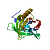 8aejC 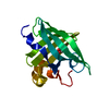 3l4rS S: Starting model for refinement C: citing same article ( |
|---|---|
| Similar structure data |
-Links
 PDBj
PDBj

- Assembly
| Deposited unit | 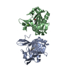
| ||||||||||||
|---|---|---|---|---|---|---|---|---|---|---|---|---|---|
| 1 | 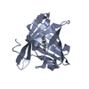
| ||||||||||||
| 2 | 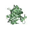
| ||||||||||||
| Unit cell |
|
-Components
| #1: Protein | Mass: 19974.135 Da / Num. of mol.: 2 Source method: isolated from a genetically manipulated source Source: (gene. exp.)  #2: Chemical |   Mass: 156.265 Da / Num. of mol.: 2 / Source method: obtained synthetically / Formula: C10H20O / Feature type: SUBJECT OF INVESTIGATION Mass: 156.265 Da / Num. of mol.: 2 / Source method: obtained synthetically / Formula: C10H20O / Feature type: SUBJECT OF INVESTIGATION#3: Chemical |   Mass: 24.305 Da / Num. of mol.: 2 / Source method: obtained synthetically / Formula: Mg Mass: 24.305 Da / Num. of mol.: 2 / Source method: obtained synthetically / Formula: Mg#4: Water | ChemComp-HOH / |  Mass: 18.015 Da / Num. of mol.: 169 / Source method: isolated from a natural source / Formula: H2O Mass: 18.015 Da / Num. of mol.: 169 / Source method: isolated from a natural source / Formula: H2OHas ligand of interest | Y | Has protein modification | Y | |
|---|
-Experimental details
-Experiment
| Experiment | Method: X-RAY DIFFRACTION / Number of used crystals: 1 |
|---|
- Sample preparation
| Crystal | Density Matthews: 2.03 Å3/Da / Density % sol: 39.55 % |
|---|---|
| Crystal grow | Temperature: 293 K / Method: vapor diffusion, sitting drop Details: 35% PEG 4000, 0.2 M MgCl2 in 0.1 M pH 8.0 Tris buffer |
-Data collection
| Diffraction | Mean temperature: 100 K / Serial crystal experiment: N |
|---|---|
| Diffraction source | Source: SYNCHROTRON / Site: SOLEIL / Beamline: PROXIMA 1 / Wavelength: 0.978566 Å |
| Detector | Type: DECTRIS EIGER X 16M / Detector: PIXEL / Date: Dec 12, 2020 |
| Radiation | Protocol: SINGLE WAVELENGTH / Monochromatic (M) / Laue (L): M / Scattering type: x-ray |
| Radiation wavelength | Wavelength: 0.978566 Å / Relative weight: 1 |
| Reflection | Resolution: 1.74→48.69 Å / Num. obs: 32275 / % possible obs: 97 % / Redundancy: 6.9 % / Biso Wilson estimate: 24.03 Å2 / CC1/2: 0.999 / Rmerge(I) obs: 0.052 / Rpim(I) all: 0.021 / Rrim(I) all: 0.056 / Net I/σ(I): 19 |
| Reflection shell | Resolution: 1.74→1.77 Å / Redundancy: 6.6 % / Rmerge(I) obs: 0.376 / Num. unique obs: 1535 / CC1/2: 0.949 / Rpim(I) all: 0.153 / Rrim(I) all: 0.407 / % possible all: 85 |
- Processing
| Software |
| ||||||||||||||||||||||||||||||||||||||||||||||||||||||||||||||||||||||||||||||||||||
|---|---|---|---|---|---|---|---|---|---|---|---|---|---|---|---|---|---|---|---|---|---|---|---|---|---|---|---|---|---|---|---|---|---|---|---|---|---|---|---|---|---|---|---|---|---|---|---|---|---|---|---|---|---|---|---|---|---|---|---|---|---|---|---|---|---|---|---|---|---|---|---|---|---|---|---|---|---|---|---|---|---|---|---|---|---|
| Refinement | Method to determine structure: MOLECULAR REPLACEMENT Starting model: 3L4R Resolution: 1.74→38.91 Å / SU ML: 0.1886 / Cross valid method: FREE R-VALUE / σ(F): 1.34 / Phase error: 24.5942 Stereochemistry target values: GeoStd + Monomer Library + CDL v1.2
| ||||||||||||||||||||||||||||||||||||||||||||||||||||||||||||||||||||||||||||||||||||
| Solvent computation | Shrinkage radii: 0.9 Å / VDW probe radii: 1.11 Å / Solvent model: FLAT BULK SOLVENT MODEL | ||||||||||||||||||||||||||||||||||||||||||||||||||||||||||||||||||||||||||||||||||||
| Displacement parameters | Biso mean: 29.9 Å2 | ||||||||||||||||||||||||||||||||||||||||||||||||||||||||||||||||||||||||||||||||||||
| Refinement step | Cycle: LAST / Resolution: 1.74→38.91 Å
| ||||||||||||||||||||||||||||||||||||||||||||||||||||||||||||||||||||||||||||||||||||
| Refine LS restraints |
| ||||||||||||||||||||||||||||||||||||||||||||||||||||||||||||||||||||||||||||||||||||
| LS refinement shell |
|
