 Movie
Movie Controller

[English] 日本語
 Yorodumi
- PDB-8ae5: Crystal structure of human legumain in complex with macrocypin 1a -
+ Open data

- Basic information
| Entry | Database: PDB / ID: 8ae5 | |||||||||
|---|---|---|---|---|---|---|---|---|---|---|
| Title | Crystal structure of human legumain in complex with macrocypin 1a | |||||||||
 Components Components |
| |||||||||
 Keywords Keywords | HYDROLASE / cysteine protease / ligase / asparaginyl endopeptidase / AEP / inhibitor / exosite / active site / substrate | |||||||||
| Function / homology |  Function and homology information Function and homology informationnegative regulation of ERBB signaling pathway / legumain / vacuolar protein processing / antigen processing and presentation of exogenous peptide antigen via MHC class I, TAP-dependent / renal system process / Vitamin D (calciferol) metabolism / vitamin D metabolic process / receptor catabolic process / endolysosome lumen / Trafficking and processing of endosomal TLR ...negative regulation of ERBB signaling pathway / legumain / vacuolar protein processing / antigen processing and presentation of exogenous peptide antigen via MHC class I, TAP-dependent / renal system process / Vitamin D (calciferol) metabolism / vitamin D metabolic process / receptor catabolic process / endolysosome lumen / Trafficking and processing of endosomal TLR / positive regulation of monocyte chemotaxis / dendritic spine organization / positive regulation of endothelial cell chemotaxis / negative regulation of multicellular organism growth / response to acidic pH / cellular response to hepatocyte growth factor stimulus / associative learning / cysteine-type endopeptidase inhibitor activity / endopeptidase activator activity / MHC class II antigen presentation / positive regulation of mitotic cell cycle / lysosomal lumen / cellular response to calcium ion / : / positive regulation of long-term synaptic potentiation / protein maturation / tau protein binding / antigen processing and presentation of exogenous peptide antigen via MHC class II / cellular response to amyloid-beta / memory / apical part of cell / late endosome / peptidase activity / negative regulation of neuron apoptotic process / lysosome / negative regulation of gene expression / cysteine-type endopeptidase activity / positive regulation of cell population proliferation / perinuclear region of cytoplasm / proteolysis / extracellular exosome / extracellular region / cytoplasm Similarity search - Function | |||||||||
| Biological species |  Homo sapiens (human) Homo sapiens (human) Macrolepiota procera (parasol mushroom) Macrolepiota procera (parasol mushroom) | |||||||||
| Method |  X-RAY DIFFRACTION / SYNCHROTRON / MOLECULAR REPLACEMENT / Resolution: 2.29 Å X-RAY DIFFRACTION / SYNCHROTRON / MOLECULAR REPLACEMENT / Resolution: 2.29 Å | |||||||||
 Authors Authors | Elamin, T. / Brandstetter, H. / Dall, E. | |||||||||
| Funding support |  Austria, 1items Austria, 1items
| |||||||||
 Citation Citation | Journal: J.Biol.Chem. / Year: 2022 Title: Structural and functional studies of legumain-mycocypin complexes revealed a competitive, exosite-regulated mode of interaction. Authors: Elamin, T. / Santos, N.P. / Briza, P. / Brandstetter, H. / Dall, E. | |||||||||
| History |
|
- Structure visualization
| Structure viewer | Molecule: MolmilJmol/JSmol |
|---|
- Downloads & links
-Download
| PDBx/mmCIF format | 8ae5.cif.gz | 436.9 KB | Display | PDBx/mmCIF format |
|---|---|---|---|---|
| PDB format | pdb8ae5.ent.gz | 302.3 KB | Display | PDB format |
| PDBx/mmJSON format | 8ae5.json.gz | Tree view | PDBx/mmJSON format | |
| Others |  Other downloads Other downloads |
-Validation report
| Arichive directory | https://data.pdbj.org/pub/pdb/validation_reports/ae/8ae5ftp://data.pdbj.org/pub/pdb/validation_reports/ae/8ae5 | HTTPS FTP |
|---|
-Related structure data
| Related structure data |  8ae4C  7o50S S: Starting model for refinement C: citing same article ( |
|---|---|
| Similar structure data |
-Links
 PDBj
PDBj


- Assembly
| Deposited unit | 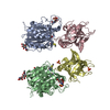
| ||||||||||||||||||||||||||||||||||||||||||||||||||||||||||||||||||||||||||||||||||||||||||||||||||||
|---|---|---|---|---|---|---|---|---|---|---|---|---|---|---|---|---|---|---|---|---|---|---|---|---|---|---|---|---|---|---|---|---|---|---|---|---|---|---|---|---|---|---|---|---|---|---|---|---|---|---|---|---|---|---|---|---|---|---|---|---|---|---|---|---|---|---|---|---|---|---|---|---|---|---|---|---|---|---|---|---|---|---|---|---|---|---|---|---|---|---|---|---|---|---|---|---|---|---|---|---|---|
| 1 | 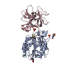
| ||||||||||||||||||||||||||||||||||||||||||||||||||||||||||||||||||||||||||||||||||||||||||||||||||||
| 2 | 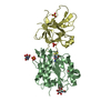
| ||||||||||||||||||||||||||||||||||||||||||||||||||||||||||||||||||||||||||||||||||||||||||||||||||||
| Unit cell |
| ||||||||||||||||||||||||||||||||||||||||||||||||||||||||||||||||||||||||||||||||||||||||||||||||||||
| Noncrystallographic symmetry (NCS) | NCS domain:
NCS domain segments:
NCS ensembles :
NCS oper:
|
-Components
-Protein , 2 types, 4 molecules ABCD
| #1: Protein | Mass: 30133.838 Da / Num. of mol.: 2 Source method: isolated from a genetically manipulated source Source: (gene. exp.) Homo sapiens (human) / Gene: LGMN, PRSC1 / Production host:  Leishmania tarentolae (eukaryote) / References: UniProt: Q99538, legumain Leishmania tarentolae (eukaryote) / References: UniProt: Q99538, legumain#2: Protein | Mass: 20283.607 Da / Num. of mol.: 2 Source method: isolated from a genetically manipulated source Source: (gene. exp.) Macrolepiota procera (parasol mushroom)Production host:  |
|---|
-Sugars , 2 types, 5 molecules 
| #3: Polysaccharide | 2-acetamido-2-deoxy-beta-D-glucopyranose-(1-4)-2-acetamido-2-deoxy-beta-D-glucopyranose |
|---|---|
| #4: Sugar | ChemComp-NAG /  Type: D-saccharide, beta linking / Mass: 221.208 Da / Num. of mol.: 4 / Source method: obtained synthetically / Formula: C8H15NO6 Type: D-saccharide, beta linking / Mass: 221.208 Da / Num. of mol.: 4 / Source method: obtained synthetically / Formula: C8H15NO6 |
-Non-polymers , 2 types, 85 molecules 

| #5: Chemical | ChemComp-SO4 /  Mass: 96.063 Da / Num. of mol.: 5 / Source method: isolated from a natural source / Formula: SO4 Mass: 96.063 Da / Num. of mol.: 5 / Source method: isolated from a natural source / Formula: SO4#6: Water | ChemComp-HOH / | Mass: 18.015 Da / Num. of mol.: 80 / Source method: isolated from a natural source / Formula: H2O |
|---|
-Details
| Has ligand of interest | N |
|---|
-Experimental details
-Experiment
| Experiment | Method: X-RAY DIFFRACTION / Number of used crystals: 1 |
|---|
- Sample preparation
| Crystal | Density Matthews: 4.01 Å3/Da / Density % sol: 69.36 % |
|---|---|
| Crystal grow | Temperature: 277 K / Method: vapor diffusion, sitting drop / Details: 1.6 M Ammonium Sulfate, 500 mM LiCl |
-Data collection
| Diffraction | Mean temperature: 100 K / Serial crystal experiment: N | ||||||||||||||||||||||||||||||||||||||||||||||||||||||||||||||||||||||||||||||||||||||||||||||||||||
|---|---|---|---|---|---|---|---|---|---|---|---|---|---|---|---|---|---|---|---|---|---|---|---|---|---|---|---|---|---|---|---|---|---|---|---|---|---|---|---|---|---|---|---|---|---|---|---|---|---|---|---|---|---|---|---|---|---|---|---|---|---|---|---|---|---|---|---|---|---|---|---|---|---|---|---|---|---|---|---|---|---|---|---|---|---|---|---|---|---|---|---|---|---|---|---|---|---|---|---|---|---|
| Diffraction source | Source: SYNCHROTRON / Site: ESRF  / Beamline: MASSIF-3 / Wavelength: 0.9677 Å / Beamline: MASSIF-3 / Wavelength: 0.9677 Å | ||||||||||||||||||||||||||||||||||||||||||||||||||||||||||||||||||||||||||||||||||||||||||||||||||||
| Detector | Type: DECTRIS EIGER X 4M / Detector: PIXEL / Date: Dec 12, 2017 | ||||||||||||||||||||||||||||||||||||||||||||||||||||||||||||||||||||||||||||||||||||||||||||||||||||
| Radiation | Protocol: SINGLE WAVELENGTH / Monochromatic (M) / Laue (L): M / Scattering type: x-ray | ||||||||||||||||||||||||||||||||||||||||||||||||||||||||||||||||||||||||||||||||||||||||||||||||||||
| Radiation wavelength | Wavelength: 0.9677 Å / Relative weight: 1 | ||||||||||||||||||||||||||||||||||||||||||||||||||||||||||||||||||||||||||||||||||||||||||||||||||||
| Reflection | Resolution: 2.28→47.39 Å / Num. obs: 70688 / % possible obs: 99.1 % / Redundancy: 11.361 % / Biso Wilson estimate: 74.8 Å2 / CC1/2: 0.999 / Rmerge(I) obs: 0.109 / Rrim(I) all: 0.114 / Χ2: 1.248 / Net I/σ(I): 13.72 / Num. measured all: 803083 | ||||||||||||||||||||||||||||||||||||||||||||||||||||||||||||||||||||||||||||||||||||||||||||||||||||
| Reflection shell | Diffraction-ID: 1
|
- Processing
| Software |
| |||||||||||||||||||||||||||||||||||||||||||||||||||||||||||||||||||||||||||||||||||||||||||||||||||||||||||||||||||||||||||||||||||||||||||||||||||||||||||||||||||||||||||||||||||||||||||||
|---|---|---|---|---|---|---|---|---|---|---|---|---|---|---|---|---|---|---|---|---|---|---|---|---|---|---|---|---|---|---|---|---|---|---|---|---|---|---|---|---|---|---|---|---|---|---|---|---|---|---|---|---|---|---|---|---|---|---|---|---|---|---|---|---|---|---|---|---|---|---|---|---|---|---|---|---|---|---|---|---|---|---|---|---|---|---|---|---|---|---|---|---|---|---|---|---|---|---|---|---|---|---|---|---|---|---|---|---|---|---|---|---|---|---|---|---|---|---|---|---|---|---|---|---|---|---|---|---|---|---|---|---|---|---|---|---|---|---|---|---|---|---|---|---|---|---|---|---|---|---|---|---|---|---|---|---|---|---|---|---|---|---|---|---|---|---|---|---|---|---|---|---|---|---|---|---|---|---|---|---|---|---|---|---|---|---|---|---|---|---|
| Refinement | Method to determine structure: MOLECULAR REPLACEMENT Starting model: 7o50 Resolution: 2.29→47.39 Å / SU ML: 0.5016 / Cross valid method: FREE R-VALUE / σ(F): 1.32 / Phase error: 34.4693 Stereochemistry target values: GeoStd + Monomer Library + CDL v1.2
| |||||||||||||||||||||||||||||||||||||||||||||||||||||||||||||||||||||||||||||||||||||||||||||||||||||||||||||||||||||||||||||||||||||||||||||||||||||||||||||||||||||||||||||||||||||||||||||
| Solvent computation | Shrinkage radii: 0.9 Å / VDW probe radii: 1.11 Å / Solvent model: FLAT BULK SOLVENT MODEL | |||||||||||||||||||||||||||||||||||||||||||||||||||||||||||||||||||||||||||||||||||||||||||||||||||||||||||||||||||||||||||||||||||||||||||||||||||||||||||||||||||||||||||||||||||||||||||||
| Displacement parameters | Biso mean: 93.69 Å2 | |||||||||||||||||||||||||||||||||||||||||||||||||||||||||||||||||||||||||||||||||||||||||||||||||||||||||||||||||||||||||||||||||||||||||||||||||||||||||||||||||||||||||||||||||||||||||||||
| Refinement step | Cycle: LAST / Resolution: 2.29→47.39 Å
| |||||||||||||||||||||||||||||||||||||||||||||||||||||||||||||||||||||||||||||||||||||||||||||||||||||||||||||||||||||||||||||||||||||||||||||||||||||||||||||||||||||||||||||||||||||||||||||
| Refine LS restraints |
| |||||||||||||||||||||||||||||||||||||||||||||||||||||||||||||||||||||||||||||||||||||||||||||||||||||||||||||||||||||||||||||||||||||||||||||||||||||||||||||||||||||||||||||||||||||||||||||
| Refine LS restraints NCS |
| |||||||||||||||||||||||||||||||||||||||||||||||||||||||||||||||||||||||||||||||||||||||||||||||||||||||||||||||||||||||||||||||||||||||||||||||||||||||||||||||||||||||||||||||||||||||||||||
| LS refinement shell |
| |||||||||||||||||||||||||||||||||||||||||||||||||||||||||||||||||||||||||||||||||||||||||||||||||||||||||||||||||||||||||||||||||||||||||||||||||||||||||||||||||||||||||||||||||||||||||||||
| Refinement TLS params. | Method: refined / Origin x: 3.23212958042 Å / Origin y: -53.7248821459 Å / Origin z: 3.40439353297 Å
| |||||||||||||||||||||||||||||||||||||||||||||||||||||||||||||||||||||||||||||||||||||||||||||||||||||||||||||||||||||||||||||||||||||||||||||||||||||||||||||||||||||||||||||||||||||||||||||
| Refinement TLS group | Selection details: all |
