 Movie
Movie Controller


+ Open data

- Basic information
| Entry | Database: PDB / ID: 8ad6 | ||||||
|---|---|---|---|---|---|---|---|
| Title | Structure of DarB bound to c-di-AMP | ||||||
 Components Components | CBS domain-containing protein | ||||||
 Keywords Keywords | SIGNALING PROTEIN / c-di-AMP / potassium stress / ppGpp / Rel / stringent factor / CbpB | ||||||
| Function / homology | CBS domain superfamily / Chem-2BA / CBS domain-containing protein Function and homology information Function and homology information | ||||||
| Biological species |  | ||||||
| Method |  X-RAY DIFFRACTION / SYNCHROTRON / MOLECULAR REPLACEMENT / Resolution: 1.52 Å X-RAY DIFFRACTION / SYNCHROTRON / MOLECULAR REPLACEMENT / Resolution: 1.52 Å | ||||||
 Authors Authors | Garcia-Pino, A. / Talavera, A. | ||||||
| Funding support |  Belgium, 1items Belgium, 1items
| ||||||
 Citation Citation | Journal: To Be Published Title: Structure of DarB bound to c-di-AMP Authors: Garcia-Pino, A. / Talavera, A. | ||||||
| History |
|
- Structure visualization
| Structure viewer | Molecule: MolmilJmol/JSmol |
|---|
- Downloads & links
-Download
| PDBx/mmCIF format | 8ad6.cif.gz | 77.2 KB | Display | PDBx/mmCIF format |
|---|---|---|---|---|
| PDB format | pdb8ad6.ent.gz | 55.4 KB | Display | PDB format |
| PDBx/mmJSON format | 8ad6.json.gz | Tree view | PDBx/mmJSON format | |
| Others |  Other downloads Other downloads |
-Validation report
| Arichive directory | https://data.pdbj.org/pub/pdb/validation_reports/ad/8ad6ftp://data.pdbj.org/pub/pdb/validation_reports/ad/8ad6 | HTTPS FTP |
|---|
-Related structure data
| Related structure data | 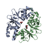 6yj8S S: Starting model for refinement |
|---|---|
| Similar structure data |
-Links
 PDBj
PDBj- Assembly
| Deposited unit | 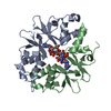
| ||||||||||||
|---|---|---|---|---|---|---|---|---|---|---|---|---|---|
| 1 |
| ||||||||||||
| Unit cell |
|
-Components
| #1: Protein | Mass: 16653.346 Da / Num. of mol.: 2 Source method: isolated from a genetically manipulated source Source: (gene. exp.) Production host: References: UniProt: A0A164SLA6 #2: Chemical | 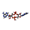  Mass: 658.412 Da / Num. of mol.: 2 / Source method: obtained synthetically / Formula: C20H24N10O12P2 / Feature type: SUBJECT OF INVESTIGATION Mass: 658.412 Da / Num. of mol.: 2 / Source method: obtained synthetically / Formula: C20H24N10O12P2 / Feature type: SUBJECT OF INVESTIGATION#3: Water | ChemComp-HOH / |  Mass: 18.015 Da / Num. of mol.: 271 / Source method: isolated from a natural source / Formula: H2O Mass: 18.015 Da / Num. of mol.: 271 / Source method: isolated from a natural source / Formula: H2OHas ligand of interest | Y | |
|---|
-Experimental details
-Experiment
| Experiment | Method: X-RAY DIFFRACTION / Number of used crystals: 1 |
|---|
- Sample preparation
| Crystal | Density Matthews: 2.18 Å3/Da / Density % sol: 43.54 % |
|---|---|
| Crystal grow | Temperature: 293.16 K / Method: vapor diffusion, sitting drop / pH: 6 Details: 0.2 M Lithium sulfate 0.1 M MES 6.0 20 % w/v PEG 4000 |
-Data collection
| Diffraction | Mean temperature: 100 K / Serial crystal experiment: N |
|---|---|
| Diffraction source | Source: SYNCHROTRON / Site: SOLEIL  / Beamline: PROXIMA 2 / Wavelength: 0.9801 Å / Beamline: PROXIMA 2 / Wavelength: 0.9801 Å |
| Detector | Type: DECTRIS EIGER X 16M / Detector: PIXEL / Date: Apr 26, 2020 |
| Radiation | Protocol: SINGLE WAVELENGTH / Monochromatic (M) / Laue (L): M / Scattering type: x-ray |
| Radiation wavelength | Wavelength: 0.9801 Å / Relative weight: 1 |
| Reflection | Resolution: 1.52→52.81 Å / Num. obs: 40317 / % possible obs: 88 % / Redundancy: 7.3 % / CC1/2: 0.999 / Rpim(I) all: 0.021 / Net I/σ(I): 19.2 |
| Reflection shell | Resolution: 1.52→1.57 Å / Num. unique obs: 998 / CC1/2: 0.826 / Rpim(I) all: 0.251 |
- Processing
| Software |
| ||||||||||||||||||||||||
|---|---|---|---|---|---|---|---|---|---|---|---|---|---|---|---|---|---|---|---|---|---|---|---|---|---|
| Refinement | Method to determine structure: MOLECULAR REPLACEMENT Starting model: 6YJ8 Resolution: 1.52→52.81 Å / Cross valid method: THROUGHOUT Stereochemistry target values: GeoStd + Monomer Library + CDL v1.2
| ||||||||||||||||||||||||
| Displacement parameters | Biso mean: 22.73 Å2 | ||||||||||||||||||||||||
| Refinement step | Cycle: LAST / Resolution: 1.52→52.81 Å
| ||||||||||||||||||||||||
| Refine LS restraints |
|
