 Movie
Movie Controller


+ Open data

- Basic information
| Entry | Database: PDB / ID: 8acv | ||||||
|---|---|---|---|---|---|---|---|
| Title | WelO5* bound to Zn(II), Cl, and 2-oxoglutarate | ||||||
 Components Components | Oxidoreductase | ||||||
 Keywords Keywords | OXIDOREDUCTASE / WelO5* 2-oxoglutarate-dependent halogenase | ||||||
| Function / homology | : / Carrier-protein-independent halogenase WelO5 / metal ion binding / ACETATE ION / 2-OXOGLUTARIC ACID / Oxidoreductase Function and homology information Function and homology information | ||||||
| Biological species |  Hapalosiphon welwitschii UH IC-52-3 (bacteria) Hapalosiphon welwitschii UH IC-52-3 (bacteria) | ||||||
| Method |  X-RAY DIFFRACTION / SYNCHROTRON / MOLECULAR REPLACEMENT / Resolution: 2.26 Å X-RAY DIFFRACTION / SYNCHROTRON / MOLECULAR REPLACEMENT / Resolution: 2.26 Å | ||||||
 Authors Authors | Buller, R. / Hueppi, S. / Voss, M. / Hayashi, T. | ||||||
| Funding support |  Switzerland, 1items Switzerland, 1items
| ||||||
 Citation Citation | Journal: Chemcatchem / Year: 2022 Title: Enzyme engineering enables inversion of substrate stereopreference of the halogenase WelO5* Authors: Voss, M. / Huppi, S. / Schaub, D. / Hayashi, T. / Ligibel, M. / Sager, E. / Schroer, K. / Snajdrova, R. / Buller, R.M.U. | ||||||
| History |
|
- Structure visualization
| Structure viewer | Molecule: MolmilJmol/JSmol |
|---|
- Downloads & links
-Download
| PDBx/mmCIF format | 8acv.cif.gz | 239.6 KB | Display | PDBx/mmCIF format |
|---|---|---|---|---|
| PDB format | pdb8acv.ent.gz | 193.1 KB | Display | PDB format |
| PDBx/mmJSON format | 8acv.json.gz | Tree view | PDBx/mmJSON format | |
| Others |  Other downloads Other downloads |
-Validation report
| Summary document | 8acv_validation.pdf.gz | 1.5 MB | Display | wwPDB validaton report |
|---|---|---|---|---|
| Full document | 8acv_full_validation.pdf.gz | 1.5 MB | Display | |
| Data in XML | 8acv_validation.xml.gz | 21.8 KB | Display | |
| Data in CIF | 8acv_validation.cif.gz | 29.9 KB | Display | |
| Arichive directory | https://data.pdbj.org/pub/pdb/validation_reports/ac/8acvftp://data.pdbj.org/pub/pdb/validation_reports/ac/8acv | HTTPS FTP |
-Related structure data
| Related structure data |  8autC 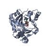 5iqsS S: Starting model for refinement C: citing same article ( |
|---|---|
| Similar structure data |
-Links
 PDBj
PDBj

- Assembly
| Deposited unit | 
| ||||||||
|---|---|---|---|---|---|---|---|---|---|
| 1 | 
| ||||||||
| 2 | 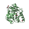
| ||||||||
| Unit cell |
| ||||||||
| Components on special symmetry positions |
|
-Components
-Protein , 1 types, 2 molecules AB
| #1: Protein | Mass: 34611.953 Da / Num. of mol.: 2 Source method: isolated from a genetically manipulated source Source: (gene. exp.) Hapalosiphon welwitschii UH IC-52-3 (bacteria)Gene: welO15 / Production host: |
|---|
-Non-polymers , 6 types, 77 molecules 


| #2: Chemical |  Mass: 146.098 Da / Num. of mol.: 2 / Source method: obtained synthetically / Formula: C5H6O5 / Feature type: SUBJECT OF INVESTIGATION Mass: 146.098 Da / Num. of mol.: 2 / Source method: obtained synthetically / Formula: C5H6O5 / Feature type: SUBJECT OF INVESTIGATION#3: Chemical |  Mass: 35.453 Da / Num. of mol.: 2 / Source method: obtained synthetically / Formula: Cl / Feature type: SUBJECT OF INVESTIGATION Mass: 35.453 Da / Num. of mol.: 2 / Source method: obtained synthetically / Formula: Cl / Feature type: SUBJECT OF INVESTIGATION#4: Chemical |  Mass: 65.409 Da / Num. of mol.: 2 / Source method: obtained synthetically / Formula: Zn / Feature type: SUBJECT OF INVESTIGATION Mass: 65.409 Da / Num. of mol.: 2 / Source method: obtained synthetically / Formula: Zn / Feature type: SUBJECT OF INVESTIGATION#5: Chemical | ChemComp-GOL /  Mass: 92.094 Da / Num. of mol.: 4 / Source method: obtained synthetically / Formula: C3H8O3 Mass: 92.094 Da / Num. of mol.: 4 / Source method: obtained synthetically / Formula: C3H8O3#6: Chemical | ChemComp-ACT /  Mass: 59.044 Da / Num. of mol.: 6 / Source method: obtained synthetically / Formula: C2H3O2 Mass: 59.044 Da / Num. of mol.: 6 / Source method: obtained synthetically / Formula: C2H3O2#7: Water | ChemComp-HOH / | Mass: 18.015 Da / Num. of mol.: 61 / Source method: isolated from a natural source / Formula: H2O |
|---|
-Details
| Has ligand of interest | Y |
|---|
-Experimental details
-Experiment
| Experiment | Method: X-RAY DIFFRACTION / Number of used crystals: 1 |
|---|
- Sample preparation
| Crystal | Density Matthews: 1.95 Å3/Da / Density % sol: 36.9 % |
|---|---|
| Crystal grow | Temperature: 293.15 K / Method: vapor diffusion, sitting drop / pH: 4.5 / Details: 22.14% PEG-4000, 30 mM sodium acetate pH 4.5 |
-Data collection
| Diffraction | Mean temperature: 100 K / Serial crystal experiment: N |
|---|---|
| Diffraction source | Source: SYNCHROTRON / Site: SLS / Beamline: X06DA / Wavelength: 1.000002 Å |
| Detector | Type: DECTRIS PILATUS 2M-F / Detector: PIXEL / Date: May 24, 2019 |
| Radiation | Protocol: SINGLE WAVELENGTH / Monochromatic (M) / Laue (L): M / Scattering type: x-ray |
| Radiation wavelength | Wavelength: 1.000002 Å / Relative weight: 1 |
| Reflection | Resolution: 2.26→47.77 Å / Num. obs: 25004 / % possible obs: 98.4 % / Redundancy: 5.4 % / Biso Wilson estimate: 58.09 Å2 / CC1/2: 0.999 / Net I/σ(I): 15.1 |
| Reflection shell | Resolution: 2.269→2.281 Å / Num. unique obs: 2283 / Rrim(I) all: 0.174 |
- Processing
| Software |
| ||||||||||||||||||||||||||||||||||||||||||||||||||||||||||||||||||||||||||||||||||||||||||||||||||||||||||||||||||
|---|---|---|---|---|---|---|---|---|---|---|---|---|---|---|---|---|---|---|---|---|---|---|---|---|---|---|---|---|---|---|---|---|---|---|---|---|---|---|---|---|---|---|---|---|---|---|---|---|---|---|---|---|---|---|---|---|---|---|---|---|---|---|---|---|---|---|---|---|---|---|---|---|---|---|---|---|---|---|---|---|---|---|---|---|---|---|---|---|---|---|---|---|---|---|---|---|---|---|---|---|---|---|---|---|---|---|---|---|---|---|---|---|---|---|---|
| Refinement | Method to determine structure: MOLECULAR REPLACEMENT Starting model: 5IQS Resolution: 2.26→47.77 Å / Cor.coef. Fo:Fc: 0.924 / Cor.coef. Fo:Fc free: 0.903 / SU R Cruickshank DPI: 0.373 / Cross valid method: THROUGHOUT / σ(F): 0 / SU R Blow DPI: 0.344 / SU Rfree Blow DPI: 0.222 / SU Rfree Cruickshank DPI: 0.23
| ||||||||||||||||||||||||||||||||||||||||||||||||||||||||||||||||||||||||||||||||||||||||||||||||||||||||||||||||||
| Displacement parameters |
| ||||||||||||||||||||||||||||||||||||||||||||||||||||||||||||||||||||||||||||||||||||||||||||||||||||||||||||||||||
| Refine analyze | Luzzati coordinate error obs: 0.34 Å | ||||||||||||||||||||||||||||||||||||||||||||||||||||||||||||||||||||||||||||||||||||||||||||||||||||||||||||||||||
| Refinement step | Cycle: 1 / Resolution: 2.26→47.77 Å
| ||||||||||||||||||||||||||||||||||||||||||||||||||||||||||||||||||||||||||||||||||||||||||||||||||||||||||||||||||
| Refine LS restraints |
| ||||||||||||||||||||||||||||||||||||||||||||||||||||||||||||||||||||||||||||||||||||||||||||||||||||||||||||||||||
| LS refinement shell | Resolution: 2.26→2.28 Å / Total num. of bins used: 50
| ||||||||||||||||||||||||||||||||||||||||||||||||||||||||||||||||||||||||||||||||||||||||||||||||||||||||||||||||||
| Refinement TLS params. | Method: refined / Refine-ID: X-RAY DIFFRACTION
| ||||||||||||||||||||||||||||||||||||||||||||||||||||||||||||||||||||||||||||||||||||||||||||||||||||||||||||||||||
| Refinement TLS group |
|
