 Movie
Movie Controller

[English] 日本語
 Yorodumi
- PDB-8aaq: Crystal structure of the carotenoid-binding protein domain from s... -
+ Open data

- Basic information
| Entry | Database: PDB / ID: 8aaq | |||||||||
|---|---|---|---|---|---|---|---|---|---|---|
| Title | Crystal structure of the carotenoid-binding protein domain from silkworm Bombyx mori (BmCBP), CRT-416 form | |||||||||
 Components Components | Carotenoid-binding protein | |||||||||
 Keywords Keywords | TRANSPORT PROTEIN / carotenoid-binding protein / carotenoid transport / CBP / STARD / START domain / Bombyx mori | |||||||||
| Function / homology |  Function and homology information Function and homology informationvesicle tethering to endoplasmic reticulum / endoplasmic reticulum-endosome membrane contact site / late endosome membrane / lysosomal membrane / lipid binding / endoplasmic reticulum membrane Similarity search - Function | |||||||||
| Biological species |  | |||||||||
| Method |  X-RAY DIFFRACTION / MOLECULAR REPLACEMENT / Resolution: 1.8 Å X-RAY DIFFRACTION / MOLECULAR REPLACEMENT / Resolution: 1.8 Å | |||||||||
 Authors Authors | Varfolomeeva, L.A. / Slonimskiy, Y.B. / Egorkin, N.A. / Minyaev, M.E. / Faletrov, Y.V. / Boyko, K.M. / Sluchanko, N.N. | |||||||||
| Funding support |  Russian Federation, 2items Russian Federation, 2items
| |||||||||
 Citation Citation | Journal: Crystallography Reports / Year: 2022 Title: Preparation and Structural Studies of the Silkworm Carotenoid-Binding Protein Complexed with a New Pigment Authors: Varfolomeeva, L.A. / Slonimskiy, Y.B. / Egorkin, N.A. / Minyaev, M.E. / Faletrov, Y.V. / Boyko, K.M. / Sluchanko, N.N. | |||||||||
| History |
|
- Structure visualization
| Structure viewer | Molecule: MolmilJmol/JSmol |
|---|
- Downloads & links
-Download
| PDBx/mmCIF format | 8aaq.cif.gz | 108.8 KB | Display | PDBx/mmCIF format |
|---|---|---|---|---|
| PDB format | pdb8aaq.ent.gz | 82.1 KB | Display | PDB format |
| PDBx/mmJSON format | 8aaq.json.gz | Tree view | PDBx/mmJSON format | |
| Others |  Other downloads Other downloads |
-Validation report
| Summary document | 8aaq_validation.pdf.gz | 430 KB | Display | wwPDB validaton report |
|---|---|---|---|---|
| Full document | 8aaq_full_validation.pdf.gz | 432.7 KB | Display | |
| Data in XML | 8aaq_validation.xml.gz | 12.4 KB | Display | |
| Data in CIF | 8aaq_validation.cif.gz | 17.8 KB | Display | |
| Arichive directory | https://data.pdbj.org/pub/pdb/validation_reports/aa/8aaqftp://data.pdbj.org/pub/pdb/validation_reports/aa/8aaq | HTTPS FTP |
-Related structure data
| Related structure data | 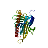 7ztqS S: Starting model for refinement |
|---|---|
| Similar structure data |
-Links
 PDBj
PDBj- Assembly
| Deposited unit | 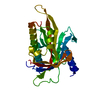
| ||||||||||||
|---|---|---|---|---|---|---|---|---|---|---|---|---|---|
| 1 |
| ||||||||||||
| Unit cell |
| ||||||||||||
| Components on special symmetry positions |
|
-Components
| #1: Protein | Mass: 28530.822 Da / Num. of mol.: 1 Source method: isolated from a genetically manipulated source Source: (gene. exp.)  |
|---|---|
| #2: Water | ChemComp-HOH /  Mass: 18.015 Da / Num. of mol.: 162 / Source method: isolated from a natural source / Formula: H2O Mass: 18.015 Da / Num. of mol.: 162 / Source method: isolated from a natural source / Formula: H2O |
-Experimental details
-Experiment
| Experiment | Method: X-RAY DIFFRACTION / Number of used crystals: 1 |
|---|
- Sample preparation
| Crystal | Density Matthews: 2.17 Å3/Da / Density % sol: 43.44 % |
|---|---|
| Crystal grow | Temperature: 288 K / Method: vapor diffusion, hanging drop Details: 0.5 M Sodium citrate tribasic dihydrate, 0.1 M BIS-TRIS propane pH 7.0 |
-Data collection
| Diffraction | Mean temperature: 100 K / Serial crystal experiment: N | ||||||||||||||||||||||||||||||
|---|---|---|---|---|---|---|---|---|---|---|---|---|---|---|---|---|---|---|---|---|---|---|---|---|---|---|---|---|---|---|---|
| Diffraction source | Source: ROTATING ANODE / Type: RIGAKU / Wavelength: 1.54184 Å | ||||||||||||||||||||||||||||||
| Detector | Type: RIGAKU HyPix-6000HE / Detector: PIXEL / Date: Feb 19, 2022 | ||||||||||||||||||||||||||||||
| Radiation | Protocol: SINGLE WAVELENGTH / Monochromatic (M) / Laue (L): M / Scattering type: x-ray | ||||||||||||||||||||||||||||||
| Radiation wavelength | Wavelength: 1.54184 Å / Relative weight: 1 | ||||||||||||||||||||||||||||||
| Reflection | Resolution: 1.8→60.13 Å / Num. obs: 22589 / % possible obs: 96 % / Redundancy: 7.6 % / CC1/2: 0.991 / Rmerge(I) obs: 0.083 / Rpim(I) all: 0.033 / Rrim(I) all: 0.09 / Net I/σ(I): 17.1 / Num. measured all: 170679 / Scaling rejects: 467 | ||||||||||||||||||||||||||||||
| Reflection shell | Diffraction-ID: 1
|
- Processing
| Software |
| |||||||||||||||||||||||||||||||||||||||||||||
|---|---|---|---|---|---|---|---|---|---|---|---|---|---|---|---|---|---|---|---|---|---|---|---|---|---|---|---|---|---|---|---|---|---|---|---|---|---|---|---|---|---|---|---|---|---|---|
| Refinement | Method to determine structure: MOLECULAR REPLACEMENT Starting model: 7ZTQ Resolution: 1.8→60.13 Å / Cor.coef. Fo:Fc: 0.949 / Cor.coef. Fo:Fc free: 0.944 / SU B: 6.284 / SU ML: 0.095 / Cross valid method: THROUGHOUT / σ(F): 0 / ESU R: 0.137 / ESU R Free: 0.13 / Stereochemistry target values: MAXIMUM LIKELIHOOD / Details: U VALUES : WITH TLS ADDED
| |||||||||||||||||||||||||||||||||||||||||||||
| Solvent computation | Ion probe radii: 0.8 Å / Shrinkage radii: 0.8 Å / VDW probe radii: 1.2 Å / Solvent model: MASK | |||||||||||||||||||||||||||||||||||||||||||||
| Displacement parameters | Biso max: 74.7 Å2 / Biso mean: 27.049 Å2 / Biso min: 13.5 Å2
| |||||||||||||||||||||||||||||||||||||||||||||
| Refinement step | Cycle: final / Resolution: 1.8→60.13 Å
| |||||||||||||||||||||||||||||||||||||||||||||
| Refine LS restraints |
| |||||||||||||||||||||||||||||||||||||||||||||
| LS refinement shell | Resolution: 1.8→1.847 Å / Rfactor Rfree error: 0 / Total num. of bins used: 20
| |||||||||||||||||||||||||||||||||||||||||||||
| Refinement TLS params. | Method: refined / Origin x: 16.0941 Å / Origin y: 12.7887 Å / Origin z: -17.2916 Å
|
