- PDB-8aaq: Crystal structure of the carotenoid-binding protein domain from s... -
+
データを開く
IDまたはキーワード:
読み込み中...
-
基本情報
登録情報
データベース: PDB / ID: 8aaq
タイトル
Crystal structure of the carotenoid-binding protein domain from silkworm Bombyx mori (BmCBP), CRT-416 form
要素
Carotenoid-binding protein
キーワード
TRANSPORT PROTEIN / carotenoid-binding protein / carotenoid transport / CBP / STARD / START domain / Bombyx mori
機能・相同性
機能・相同性情報
vesicle tethering to endoplasmic reticulum / endoplasmic reticulum-endosome membrane contact site / late endosome membrane / lysosomal membrane / lipid binding / endoplasmic reticulum membrane 類似検索 - 分子機能
: / in StAR and phosphatidylcholine transfer protein / START domain / START domain / START domain profile. / START-like domain superfamily 類似検索 - ドメイン・相同性
 ムービー
ムービー コントローラー


 データを開く
 基本情報
基本情報 要素
要素 キーワード
キーワード 機能・相同性情報
 X線回折 /
  データ登録者
 ロシア, 2件
  引用
引用 構造の表示
構造の表示 ダウンロードとリンク
 その他のダウンロード
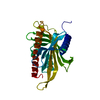
 PDBj
PDBj 集合体
集合体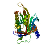

 分子量: 18.015 Da / 分子数: 162 / 由来タイプ: 天然 / 式: H2O
 試料調製
試料調製 解析
解析