 Movie
Movie Controller

[English] 日本語
 Yorodumi
- PDB-8aai: Crystal structure of the PDZ tandem of syntenin in complex with f... -
+ Open data

- Basic information
| Entry | Database: PDB / ID: 8aai | ||||||
|---|---|---|---|---|---|---|---|
| Title | Crystal structure of the PDZ tandem of syntenin in complex with fragment E5 | ||||||
 Components Components | Syntenin-1 | ||||||
 Keywords Keywords | SIGNALING PROTEIN / signaling protein cell adhesion PDZ domain syntenin syndecan drug design | ||||||
| Function / homology |  Function and homology information Function and homology informationinterleukin-5 receptor complex / interleukin-5 receptor binding / positive regulation of extracellular exosome assembly / Neurofascin interactions / syndecan binding / cytoskeletal anchor activity / substrate-dependent cell migration, cell extension / positive regulation of exosomal secretion / negative regulation of receptor internalization / frizzled binding ...interleukin-5 receptor complex / interleukin-5 receptor binding / positive regulation of extracellular exosome assembly / Neurofascin interactions / syndecan binding / cytoskeletal anchor activity / substrate-dependent cell migration, cell extension / positive regulation of exosomal secretion / negative regulation of receptor internalization / frizzled binding / Ephrin signaling / protein targeting to membrane / RIPK1-mediated regulated necrosis / positive regulation of transforming growth factor beta receptor signaling pathway / positive regulation of phosphorylation / positive regulation of epithelial to mesenchymal transition / phosphatidylinositol-4,5-bisphosphate binding / protein sequestering activity / regulation of mitotic cell cycle / adherens junction / positive regulation of JNK cascade / Regulation of necroptotic cell death / azurophil granule lumen / melanosome / extracellular vesicle / positive regulation of cell growth / actin cytoskeleton organization / blood microparticle / nuclear membrane / chemical synaptic transmission / Ras protein signal transduction / cytoskeleton / intracellular signal transduction / positive regulation of cell migration / membrane raft / protein heterodimerization activity / focal adhesion / positive regulation of cell population proliferation / Neutrophil degranulation / synapse / endoplasmic reticulum membrane / negative regulation of transcription by RNA polymerase II / extracellular space / extracellular exosome / extracellular region / nucleoplasm / identical protein binding / membrane / nucleus / plasma membrane / cytoplasm / cytosol Similarity search - Function | ||||||
| Biological species |  Homo sapiens (human) Homo sapiens (human) | ||||||
| Method |  X-RAY DIFFRACTION / SYNCHROTRON / MOLECULAR REPLACEMENT / Resolution: 2.76 Å X-RAY DIFFRACTION / SYNCHROTRON / MOLECULAR REPLACEMENT / Resolution: 2.76 Å | ||||||
 Authors Authors | Feracci, M. / Barral, K. | ||||||
| Funding support |  France, 1items France, 1items
| ||||||
 Citation Citation | Journal: J.Med.Chem. / Year: 2023 Title: Discovery of a PDZ Domain Inhibitor Targeting the Syndecan/Syntenin Protein-Protein Interaction: A Semi-Automated "Hit Identification-to-Optimization" Approach. Authors: Hoffer, L. / Garcia, M. / Leblanc, R. / Feracci, M. / Betzi, S. / Ben Yaala, K. / Daulat, A.M. / Zimmermann, P. / Roche, P. / Barral, K. / Morelli, X. | ||||||
| History |
|
- Structure visualization
| Structure viewer | Molecule: MolmilJmol/JSmol |
|---|
- Downloads & links
-Download
| PDBx/mmCIF format | 8aai.cif.gz | 261.3 KB | Display | PDBx/mmCIF format |
|---|---|---|---|---|
| PDB format | pdb8aai.ent.gz | 206.5 KB | Display | PDB format |
| PDBx/mmJSON format | 8aai.json.gz | Tree view | PDBx/mmJSON format | |
| Others |  Other downloads Other downloads |
-Validation report
| Arichive directory | https://data.pdbj.org/pub/pdb/validation_reports/aa/8aaiftp://data.pdbj.org/pub/pdb/validation_reports/aa/8aai | HTTPS FTP |
|---|
-Related structure data
| Related structure data | 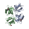 8aakC 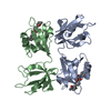 8aaoC 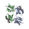 8aapC  1n99S S: Starting model for refinement C: citing same article ( |
|---|---|
| Similar structure data |
-Links
 PDBj
PDBj


- Assembly
| Deposited unit | 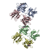
| ||||||||||||||||||||||||||||||||||||||||||||||||||||||||||||||||||||||||||||||||||||||||||||
|---|---|---|---|---|---|---|---|---|---|---|---|---|---|---|---|---|---|---|---|---|---|---|---|---|---|---|---|---|---|---|---|---|---|---|---|---|---|---|---|---|---|---|---|---|---|---|---|---|---|---|---|---|---|---|---|---|---|---|---|---|---|---|---|---|---|---|---|---|---|---|---|---|---|---|---|---|---|---|---|---|---|---|---|---|---|---|---|---|---|---|---|---|---|
| 1 | 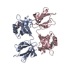
| ||||||||||||||||||||||||||||||||||||||||||||||||||||||||||||||||||||||||||||||||||||||||||||
| 2 | 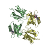
| ||||||||||||||||||||||||||||||||||||||||||||||||||||||||||||||||||||||||||||||||||||||||||||
| Unit cell |
| ||||||||||||||||||||||||||||||||||||||||||||||||||||||||||||||||||||||||||||||||||||||||||||
| Noncrystallographic symmetry (NCS) | NCS domain:
NCS domain segments: Beg auth comp-ID: ASP / Beg label comp-ID: ASP / End auth comp-ID: ALA / End label comp-ID: ALA / Refine code: 1 / Auth asym-ID: A / Label asym-ID: A / Auth seq-ID: 111 - 272 / Label seq-ID: 4 - 165
NCS ensembles :
|
-Components
| #1: Protein | Mass: 18018.688 Da / Num. of mol.: 4 Source method: isolated from a genetically manipulated source Source: (gene. exp.) Homo sapiens (human) / Gene: SDCBP, MDA9, SYCL / Production host:  #2: Chemical | ChemComp-LKV / ( 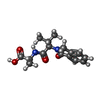  Mass: 304.341 Da / Num. of mol.: 4 / Source method: obtained synthetically / Formula: C16H20N2O4 / Feature type: SUBJECT OF INVESTIGATION Mass: 304.341 Da / Num. of mol.: 4 / Source method: obtained synthetically / Formula: C16H20N2O4 / Feature type: SUBJECT OF INVESTIGATION#3: Water | ChemComp-HOH / |  Mass: 18.015 Da / Num. of mol.: 188 / Source method: isolated from a natural source / Formula: H2O Mass: 18.015 Da / Num. of mol.: 188 / Source method: isolated from a natural source / Formula: H2OHas ligand of interest | Y | |
|---|
-Experimental details
-Experiment
| Experiment | Method: X-RAY DIFFRACTION / Number of used crystals: 1 |
|---|
- Sample preparation
| Crystal | Density Matthews: 2.25 Å3/Da / Density % sol: 45.23 % |
|---|---|
| Crystal grow | Temperature: 297 K / Method: vapor diffusion, sitting drop Details: 100mM Sodium Acetate 200mM Ammonium acetate 20% PEG 3350 PH range: 4.4 - 4.8 |
-Data collection
| Diffraction | Mean temperature: 100 K / Serial crystal experiment: N |
|---|---|
| Diffraction source | Source: SYNCHROTRON / Site: ESRF / Beamline: ID23-1 / Wavelength: 0.97625 Å |
| Detector | Type: DECTRIS PILATUS 6M / Detector: PIXEL / Date: Nov 19, 2017 |
| Radiation | Protocol: SINGLE WAVELENGTH / Monochromatic (M) / Laue (L): M / Scattering type: x-ray |
| Radiation wavelength | Wavelength: 0.97625 Å / Relative weight: 1 |
| Reflection | Resolution: 2.76→27.63 Å / Num. obs: 15909 / % possible obs: 97.29 % / Redundancy: 5.27 % / Rmerge(I) obs: 0.1388 / Rpim(I) all: 0.08908 / Net I/σ(I): 5.27 |
| Reflection shell | Resolution: 2.76→2.859 Å / Rmerge(I) obs: 0.4417 / Mean I/σ(I) obs: 2.26 / Num. unique obs: 1613 / Rpim(I) all: 0.288 / % possible all: 96.74 |
- Processing
| Software |
| ||||||||||||||||||||||||||||||||||||||||||||||||||||||||||||||||||||||||||||||||||||||||||||||||||||||||||||||||||||||||||||||||||||||||||||||||||||||||||||||||||||||||||||||||||||||
|---|---|---|---|---|---|---|---|---|---|---|---|---|---|---|---|---|---|---|---|---|---|---|---|---|---|---|---|---|---|---|---|---|---|---|---|---|---|---|---|---|---|---|---|---|---|---|---|---|---|---|---|---|---|---|---|---|---|---|---|---|---|---|---|---|---|---|---|---|---|---|---|---|---|---|---|---|---|---|---|---|---|---|---|---|---|---|---|---|---|---|---|---|---|---|---|---|---|---|---|---|---|---|---|---|---|---|---|---|---|---|---|---|---|---|---|---|---|---|---|---|---|---|---|---|---|---|---|---|---|---|---|---|---|---|---|---|---|---|---|---|---|---|---|---|---|---|---|---|---|---|---|---|---|---|---|---|---|---|---|---|---|---|---|---|---|---|---|---|---|---|---|---|---|---|---|---|---|---|---|---|---|---|---|
| Refinement | Method to determine structure: MOLECULAR REPLACEMENT Starting model: 1N99 Resolution: 2.76→27.63 Å / Cor.coef. Fo:Fc: 0.895 / Cor.coef. Fo:Fc free: 0.871 / SU B: 26.151 / SU ML: 0.507 / Cross valid method: FREE R-VALUE / ESU R Free: 0.528 Details: Hydrogens have been added in their riding positions
| ||||||||||||||||||||||||||||||||||||||||||||||||||||||||||||||||||||||||||||||||||||||||||||||||||||||||||||||||||||||||||||||||||||||||||||||||||||||||||||||||||||||||||||||||||||||
| Solvent computation | Ion probe radii: 0.8 Å / Shrinkage radii: 0.8 Å / VDW probe radii: 1.2 Å / Solvent model: MASK BULK SOLVENT | ||||||||||||||||||||||||||||||||||||||||||||||||||||||||||||||||||||||||||||||||||||||||||||||||||||||||||||||||||||||||||||||||||||||||||||||||||||||||||||||||||||||||||||||||||||||
| Displacement parameters | Biso mean: 50.548 Å2
| ||||||||||||||||||||||||||||||||||||||||||||||||||||||||||||||||||||||||||||||||||||||||||||||||||||||||||||||||||||||||||||||||||||||||||||||||||||||||||||||||||||||||||||||||||||||
| Refinement step | Cycle: LAST / Resolution: 2.76→27.63 Å
| ||||||||||||||||||||||||||||||||||||||||||||||||||||||||||||||||||||||||||||||||||||||||||||||||||||||||||||||||||||||||||||||||||||||||||||||||||||||||||||||||||||||||||||||||||||||
| Refine LS restraints |
|