 Movie
Movie Controller


+ Open data

- Basic information
| Entry | Database: PDB / ID: 8aab | ||||||
|---|---|---|---|---|---|---|---|
| Title | S148F mutant of blue-to-red fluorescent timer mRubyFT | ||||||
 Components Components | mRubyFT S148F mutant of blue-to-red fluorescent timer | ||||||
 Keywords Keywords | FLUORESCENT PROTEIN / mRubyFT / S148I mutant / flourescent timer | ||||||
| Function / homology | Green fluorescent protein-related / Green fluorescent protein / Green fluorescent protein / bioluminescence / Red fluorescent protein eqFP611 Function and homology information Function and homology information | ||||||
| Biological species |  Entacmaea quadricolor (sea anemone) Entacmaea quadricolor (sea anemone) | ||||||
| Method |  X-RAY DIFFRACTION / SYNCHROTRON / MOLECULAR REPLACEMENT / molecular replacement / Resolution: 1.6 Å X-RAY DIFFRACTION / SYNCHROTRON / MOLECULAR REPLACEMENT / molecular replacement / Resolution: 1.6 Å | ||||||
 Authors Authors | Boyko, K.M. / Nikolaeva, A.Y. / Vlaskina, A.V. / Dorovatovskii, P.V. / Subach, O.M. / Popov, V.O. / Subach, F.V. | ||||||
| Funding support |  Russian Federation, 1items Russian Federation, 1items
| ||||||
 Citation Citation | Journal: Crystallography Reports / Year: 2022 Title: mRubyFT/S147I, a mutant of blue-to-red fluorescent timer Authors: Boyko, K.M. / Nikolaeva, A.Y. / Dorovatovskii, P.V. / Vlaskina, A.V. / Subach, O.M. / Subach, F.V. | ||||||
| History |
|
- Structure visualization
| Structure viewer | Molecule: MolmilJmol/JSmol |
|---|
- Downloads & links
-Download
| PDBx/mmCIF format | 8aab.cif.gz | 103.4 KB | Display | PDBx/mmCIF format |
|---|---|---|---|---|
| PDB format | pdb8aab.ent.gz | 78.1 KB | Display | PDB format |
| PDBx/mmJSON format | 8aab.json.gz | Tree view | PDBx/mmJSON format | |
| Others |  Other downloads Other downloads |
-Validation report
| Arichive directory | https://data.pdbj.org/pub/pdb/validation_reports/aa/8aabftp://data.pdbj.org/pub/pdb/validation_reports/aa/8aab | HTTPS FTP |
|---|
-Related structure data
| Related structure data |  7qgkS S: Starting model for refinement |
|---|---|
| Similar structure data |
-Links
 PDBj
PDBj

- Assembly
| Deposited unit | 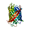
| ||||||||
|---|---|---|---|---|---|---|---|---|---|
| 1 |
| ||||||||
| Unit cell |
|
-Components
| #1: Protein | Mass: 27098.084 Da / Num. of mol.: 1 / Mutation: S148F Source method: isolated from a genetically manipulated source Source: (gene. exp.) Entacmaea quadricolor (sea anemone) / Production host:  |
|---|---|
| #2: Water | ChemComp-HOH /  Mass: 18.015 Da / Num. of mol.: 123 / Source method: isolated from a natural source / Formula: H2O Mass: 18.015 Da / Num. of mol.: 123 / Source method: isolated from a natural source / Formula: H2O |
-Experimental details
-Experiment
| Experiment | Method: X-RAY DIFFRACTION / Number of used crystals: 1 |
|---|
- Sample preparation
| Crystal | Density Matthews: 1.87 Å3/Da / Density % sol: 34.05 % |
|---|---|
| Crystal grow | Temperature: 288 K / Method: vapor diffusion, hanging drop / Details: 250 mM MgCl2; 100 mM HEPES 20% PEG 3350, pH 7,5 |
-Data collection
| Diffraction | Mean temperature: 100 K / Serial crystal experiment: N | ||||||||||||||||||||||||||||||
|---|---|---|---|---|---|---|---|---|---|---|---|---|---|---|---|---|---|---|---|---|---|---|---|---|---|---|---|---|---|---|---|
| Diffraction source | Source: SYNCHROTRON / Site: KURCHATOV SNC / Beamline: K4.4 / Wavelength: 0.79373 Å | ||||||||||||||||||||||||||||||
| Detector | Type: MAR CCD 165 mm / Detector: CCD / Date: Mar 27, 2022 | ||||||||||||||||||||||||||||||
| Radiation | Protocol: SINGLE WAVELENGTH / Monochromatic (M) / Laue (L): M / Scattering type: x-ray | ||||||||||||||||||||||||||||||
| Radiation wavelength | Wavelength: 0.79373 Å / Relative weight: 1 | ||||||||||||||||||||||||||||||
| Reflection | Resolution: 1.55→31.42 Å / Num. obs: 30195 / % possible obs: 99.7 % / Redundancy: 4.9 % / CC1/2: 0.995 / Rmerge(I) obs: 0.127 / Rpim(I) all: 0.064 / Rrim(I) all: 0.143 / Net I/σ(I): 7 / Num. measured all: 149362 / Scaling rejects: 169 | ||||||||||||||||||||||||||||||
| Reflection shell | Diffraction-ID: 1
|
-Phasing
| Phasing | Method: molecular replacement |
|---|
- Processing
| Software |
| |||||||||||||||||||||||||||||||||||||||||||||||||||||||
|---|---|---|---|---|---|---|---|---|---|---|---|---|---|---|---|---|---|---|---|---|---|---|---|---|---|---|---|---|---|---|---|---|---|---|---|---|---|---|---|---|---|---|---|---|---|---|---|---|---|---|---|---|---|---|---|---|
| Refinement | Method to determine structure: MOLECULAR REPLACEMENT Starting model: 7QGK Resolution: 1.6→31.42 Å / Cor.coef. Fo:Fc: 0.956 / Cor.coef. Fo:Fc free: 0.942 / SU B: 4.818 / SU ML: 0.081 / SU R Cruickshank DPI: 0.1007 / Cross valid method: THROUGHOUT / σ(F): 0 / ESU R: 0.101 / ESU R Free: 0.097 / Stereochemistry target values: MAXIMUM LIKELIHOOD Details: HYDROGENS HAVE BEEN ADDED IN THE RIDING POSITIONS U VALUES : WITH TLS ADDED
| |||||||||||||||||||||||||||||||||||||||||||||||||||||||
| Solvent computation | Ion probe radii: 0.8 Å / Shrinkage radii: 0.8 Å / VDW probe radii: 1.2 Å / Solvent model: MASK | |||||||||||||||||||||||||||||||||||||||||||||||||||||||
| Displacement parameters | Biso max: 51.34 Å2 / Biso mean: 16.126 Å2 / Biso min: 8.11 Å2
| |||||||||||||||||||||||||||||||||||||||||||||||||||||||
| Refinement step | Cycle: final / Resolution: 1.6→31.42 Å
| |||||||||||||||||||||||||||||||||||||||||||||||||||||||
| Refine LS restraints |
| |||||||||||||||||||||||||||||||||||||||||||||||||||||||
| LS refinement shell | Resolution: 1.6→1.641 Å / Rfactor Rfree error: 0 / Total num. of bins used: 20
| |||||||||||||||||||||||||||||||||||||||||||||||||||||||
| Refinement TLS params. | Method: refined / Origin x: 29.7536 Å / Origin y: 57.7583 Å / Origin z: 21.9898 Å
|
