 Movie
Movie Controller

[English] 日本語
 Yorodumi
- PDB-8aa5: Cryo-EM structure of the strand transfer complex of the TnsB tran... -
+ Open data

- Basic information
| Entry | Database: PDB / ID: 8aa5 | |||||||||||||||
|---|---|---|---|---|---|---|---|---|---|---|---|---|---|---|---|---|
| Title | Cryo-EM structure of the strand transfer complex of the TnsB transposase (type V-K CRISPR-associated transposon) | |||||||||||||||
 Components Components |
| |||||||||||||||
 Keywords Keywords | DNA BINDING PROTEIN / Transposase / complex / CRISPR / transposition / DNA cleavage / ligation | |||||||||||||||
| Function / homology | DNA / DNA (> 10) Function and homology information Function and homology information | |||||||||||||||
| Biological species |  Scytonema hofmannii (bacteria) Scytonema hofmannii (bacteria) | |||||||||||||||
| Method | ELECTRON MICROSCOPY / single particle reconstruction / cryo EM / Resolution: 2.46 Å | |||||||||||||||
 Authors Authors | Tenjo-Castano, F. / Sofos, N. / Lopez-Mendez, B. / Stutzke, L.S. / Fuglsang, A. / Stella, S. / Montoya, G. | |||||||||||||||
| Funding support |  Denmark, 4items Denmark, 4items
| |||||||||||||||
 Citation Citation | Journal: Nat Commun / Year: 2022 Title: Structure of the TnsB transposase-DNA complex of type V-K CRISPR-associated transposon. Authors: Francisco Tenjo-Castaño / Nicholas Sofos / Blanca López-Méndez / Luisa S Stutzke / Anders Fuglsang / Stefano Stella / Guillermo Montoya / Abstract: CRISPR-associated transposons (CASTs) are mobile genetic elements that co-opted CRISPR-Cas systems for RNA-guided transposition. Here we present the 2.4 Å cryo-EM structure of the Scytonema ...CRISPR-associated transposons (CASTs) are mobile genetic elements that co-opted CRISPR-Cas systems for RNA-guided transposition. Here we present the 2.4 Å cryo-EM structure of the Scytonema hofmannii (sh) TnsB transposase from Type V-K CAST, bound to the strand transfer DNA. The strand transfer complex displays an intertwined pseudo-symmetrical architecture. Two protomers involved in strand transfer display a catalytically competent active site composed by DDE residues, while other two, which play a key structural role, show active sites where the catalytic residues are not properly positioned for phosphodiester hydrolysis. Transposon end recognition is accomplished by the NTD1/2 helical domains. A singular in trans association of NTD1 domains of the catalytically competent subunits with the inactive DDE domains reinforces the assembly. Collectively, the structural features suggest that catalysis is coupled to protein-DNA assembly to secure proper DNA integration. DNA binding residue mutants reveal that lack of specificity decreases activity, but it could increase transposition in some cases. Our structure sheds light on the strand transfer reaction of DDE transposases and offers new insights into CAST transposition. | |||||||||||||||
| History |
|
- Structure visualization
| Structure viewer | Molecule: MolmilJmol/JSmol |
|---|
- Downloads & links
-Download
| PDBx/mmCIF format | 8aa5.cif.gz | 436.7 KB | Display | PDBx/mmCIF format |
|---|---|---|---|---|
| PDB format | pdb8aa5.ent.gz | Display | PDB format | |
| PDBx/mmJSON format | 8aa5.json.gz | Tree view | PDBx/mmJSON format | |
| Others |  Other downloads Other downloads |
-Validation report
| Arichive directory | https://data.pdbj.org/pub/pdb/validation_reports/aa/8aa5ftp://data.pdbj.org/pub/pdb/validation_reports/aa/8aa5 | HTTPS FTP |
|---|
-Related structure data
| Related structure data |  15294MC M: map data used to model this data C: citing same article ( |
|---|---|
| Similar structure data |

-Links
 PDBj
PDBj


- Assembly
| Deposited unit | 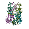
|
|---|---|
| 1 |
|
-Components
-DNA chain , 6 types, 6 molecules IJKLMN
| #2: DNA chain | Mass: 24402.717 Da / Num. of mol.: 1 / Source method: obtained synthetically / Source: (synth.) Scytonema hofmannii (bacteria) |
|---|---|
| #3: DNA chain | Mass: 22876.809 Da / Num. of mol.: 1 / Source method: obtained synthetically / Source: (synth.) Scytonema hofmannii (bacteria) |
| #4: DNA chain | Mass: 4559.971 Da / Num. of mol.: 1 / Source method: obtained synthetically / Source: (synth.) Scytonema hofmannii (bacteria) |
| #5: DNA chain | Mass: 24634.830 Da / Num. of mol.: 1 / Source method: obtained synthetically / Source: (synth.) Scytonema hofmannii (bacteria) |
| #6: DNA chain | Mass: 23238.008 Da / Num. of mol.: 1 / Source method: obtained synthetically / Source: (synth.) Scytonema hofmannii (bacteria) |
| #7: DNA chain | Mass: 4546.990 Da / Num. of mol.: 1 / Source method: obtained synthetically / Source: (synth.) Scytonema hofmannii (bacteria) |
-Protein / Non-polymers , 2 types, 6 molecules AP1BP1CP1DP1
| #1: Protein | Mass: 68094.609 Da / Num. of mol.: 4 Source method: isolated from a genetically manipulated source Source: (gene. exp.) Scytonema hofmannii (bacteria) / Production host: #8: Water | ChemComp-HOH / | Mass: 18.015 Da / Num. of mol.: 2 / Source method: isolated from a natural source / Formula: H2O |
|---|
-Experimental details
-Experiment
| Experiment | Method: ELECTRON MICROSCOPY |
|---|---|
| EM experiment | Aggregation state: PARTICLE / 3D reconstruction method: single particle reconstruction |
- Sample preparation
| Component | Name: Ternary complex of ShTnsB transposase with Strand Transfer Complex DNA. Type: COMPLEX / Entity ID: #1-#7 / Source: RECOMBINANT | |||||||||||||||||||||||||
|---|---|---|---|---|---|---|---|---|---|---|---|---|---|---|---|---|---|---|---|---|---|---|---|---|---|---|
| Molecular weight | Value: 0.359 MDa / Experimental value: YES | |||||||||||||||||||||||||
| Source (natural) | Organism: Scytonema hofmannii (bacteria) | |||||||||||||||||||||||||
| Source (recombinant) | Organism: | |||||||||||||||||||||||||
| Buffer solution | pH: 7.5 | |||||||||||||||||||||||||
| Buffer component |
| |||||||||||||||||||||||||
| Specimen | Conc.: 3.8 mg/ml / Embedding applied: NO / Shadowing applied: NO / Staining applied: NO / Vitrification applied: YES | |||||||||||||||||||||||||
| Specimen support | Grid material: GOLD / Grid mesh size: 300 divisions/in. / Grid type: UltrAuFoil R1.2/1.3 | |||||||||||||||||||||||||
| Vitrification | Instrument: FEI VITROBOT MARK IV / Cryogen name: ETHANE / Humidity: 100 % / Chamber temperature: 277 K |
- Electron microscopy imaging
| Experimental equipment |  Model: Titan Krios / Image courtesy: FEI Company |
|---|---|
| Microscopy | Model: TFS KRIOS |
| Electron gun | Electron source:  FIELD EMISSION GUN / Accelerating voltage: 300 kV / Illumination mode: FLOOD BEAM FIELD EMISSION GUN / Accelerating voltage: 300 kV / Illumination mode: FLOOD BEAM |
| Electron lens | Mode: BRIGHT FIELD / Nominal magnification: 96000 X / Nominal defocus max: 2000 nm / Nominal defocus min: 800 nm / Cs: 2.7 mm / C2 aperture diameter: 70 µm / Alignment procedure: COMA FREE |
| Specimen holder | Cryogen: NITROGEN / Specimen holder model: FEI TITAN KRIOS AUTOGRID HOLDER |
| Image recording | Average exposure time: 40 sec. / Electron dose: 40 e/Å2 / Detector mode: COUNTING / Film or detector model: FEI FALCON III (4k x 4k) / Num. of grids imaged: 1 / Num. of real images: 4728 |
- Processing
| Software |
| ||||||||||||||||||||||||||||||||||||||||||||
|---|---|---|---|---|---|---|---|---|---|---|---|---|---|---|---|---|---|---|---|---|---|---|---|---|---|---|---|---|---|---|---|---|---|---|---|---|---|---|---|---|---|---|---|---|---|
| EM software |
| ||||||||||||||||||||||||||||||||||||||||||||
| CTF correction | Type: PHASE FLIPPING AND AMPLITUDE CORRECTION | ||||||||||||||||||||||||||||||||||||||||||||
| Particle selection | Num. of particles selected: 9646000 | ||||||||||||||||||||||||||||||||||||||||||||
| Symmetry | Point symmetry: C1 (asymmetric) | ||||||||||||||||||||||||||||||||||||||||||||
| 3D reconstruction | Resolution: 2.46 Å / Resolution method: FSC 0.143 CUT-OFF / Num. of particles: 208000 / Num. of class averages: 1 / Symmetry type: POINT | ||||||||||||||||||||||||||||||||||||||||||||
| Atomic model building | Protocol: AB INITIO MODEL / Space: REAL | ||||||||||||||||||||||||||||||||||||||||||||
| Refinement | Cross valid method: NONE Stereochemistry target values: GeoStd + Monomer Library + CDL v1.2 | ||||||||||||||||||||||||||||||||||||||||||||
| Displacement parameters | Biso mean: 50.8 Å2 | ||||||||||||||||||||||||||||||||||||||||||||
| Refine LS restraints |
|
