 Movie
Movie Controller


+ Open data

- Basic information
| Entry | Database: PDB / ID: 8a90 | ||||||
|---|---|---|---|---|---|---|---|
| Title | Crystal structure of FrsH | ||||||
 Components Components | Non-heme diiron monooxygenase | ||||||
 Keywords Keywords | OXIDOREDUCTASE / NRPS | ||||||
| Function / homology |  Function and homology information Function and homology information | ||||||
| Biological species |  Chromobacterium vaccinii (bacteria) Chromobacterium vaccinii (bacteria) | ||||||
| Method |  X-RAY DIFFRACTION / SYNCHROTRON / MOLECULAR REPLACEMENT / Resolution: 2.574 Å X-RAY DIFFRACTION / SYNCHROTRON / MOLECULAR REPLACEMENT / Resolution: 2.574 Å | ||||||
 Authors Authors | Schneberger, N. / Wirtz, D.A. / Cruesemann, M. / Hagelueken, G. | ||||||
| Funding support | 1items
| ||||||
 Citation Citation | Journal: Acs Chem.Biol. / Year: 2023 Title: Adenylation Domain-Guided Recruitment of Trans- Acting Nonheme Monooxygenases in Nonribosomal Peptide Biosynthesis. Authors: Wirtz, D.A. / Schneberger, N. / Kloppel, S. / Richarz, R. / Geyer, M. / Konig, G.M. / Hagelueken, G. / Crusemann, M. | ||||||
| History |
|
- Structure visualization
| Structure viewer | Molecule: MolmilJmol/JSmol |
|---|
- Downloads & links
-Download
| PDBx/mmCIF format | 8a90.cif.gz | 271.9 KB | Display | PDBx/mmCIF format |
|---|---|---|---|---|
| PDB format | pdb8a90.ent.gz | 177.2 KB | Display | PDB format |
| PDBx/mmJSON format | 8a90.json.gz | Tree view | PDBx/mmJSON format | |
| Others |  Other downloads Other downloads |
-Validation report
| Summary document | 8a90_validation.pdf.gz | 2 MB | Display | wwPDB validaton report |
|---|---|---|---|---|
| Full document | 8a90_full_validation.pdf.gz | 2 MB | Display | |
| Data in XML | 8a90_validation.xml.gz | 44.7 KB | Display | |
| Data in CIF | 8a90_validation.cif.gz | 57.8 KB | Display | |
| Arichive directory | https://data.pdbj.org/pub/pdb/validation_reports/a9/8a90ftp://data.pdbj.org/pub/pdb/validation_reports/a9/8a90 | HTTPS FTP |
-Related structure data
| Related structure data | 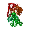 4jo0S S: Starting model for refinement |
|---|---|
| Similar structure data |
-Links
 PDBj
PDBj

- Assembly
| Deposited unit | 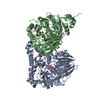
| ||||||||||||||||||
|---|---|---|---|---|---|---|---|---|---|---|---|---|---|---|---|---|---|---|---|
| 1 |
| ||||||||||||||||||
| Unit cell |
| ||||||||||||||||||
| Components on special symmetry positions |
| ||||||||||||||||||
| Noncrystallographic symmetry (NCS) | NCS domain:
NCS domain segments: Component-ID: 1 / Ens-ID: ens_1 / Beg auth comp-ID: SER / Beg label comp-ID: SER / End auth comp-ID: LEU / End label comp-ID: LEU / Auth seq-ID: 4 - 531 / Label seq-ID: 4 - 531
NCS oper: (Code: givenMatrix: (-0.346109814363, -0.82662189853, 0.443738924676), (-0.835612433918, 0.0565613263848, -0.546399740702), (0.426567528867, -0.559908075695, -0.710311966735)Vector: 74. ...NCS oper: (Code: given Matrix: (-0.346109814363, -0.82662189853, 0.443738924676), Vector: |
-Components
-Protein , 1 types, 2 molecules AB
| #1: Protein | Mass: 60273.219 Da / Num. of mol.: 2 Source method: isolated from a genetically manipulated source Source: (gene. exp.) Chromobacterium vaccinii (bacteria) / Gene: frsH / Production host: |
|---|
-Non-polymers , 5 types, 66 molecules 


| #2: Chemical |  Mass: 59.044 Da / Num. of mol.: 2 / Source method: obtained synthetically / Formula: C2H3O2 Mass: 59.044 Da / Num. of mol.: 2 / Source method: obtained synthetically / Formula: C2H3O2#3: Chemical |  Mass: 92.094 Da / Num. of mol.: 3 / Source method: obtained synthetically / Formula: C3H8O3 Mass: 92.094 Da / Num. of mol.: 3 / Source method: obtained synthetically / Formula: C3H8O3#4: Chemical |  Mass: 15.999 Da / Num. of mol.: 2 / Source method: obtained synthetically / Formula: O Mass: 15.999 Da / Num. of mol.: 2 / Source method: obtained synthetically / Formula: O#5: Chemical | ChemComp-FE /  Mass: 55.845 Da / Num. of mol.: 4 / Source method: obtained synthetically / Formula: Fe / Feature type: SUBJECT OF INVESTIGATION Mass: 55.845 Da / Num. of mol.: 4 / Source method: obtained synthetically / Formula: Fe / Feature type: SUBJECT OF INVESTIGATION#6: Water | ChemComp-HOH / | Mass: 18.015 Da / Num. of mol.: 55 / Source method: isolated from a natural source / Formula: H2O |
|---|
-Details
| Has ligand of interest | Y |
|---|---|
| Has protein modification | N |
-Experimental details
-Experiment
| Experiment | Method: X-RAY DIFFRACTION / Number of used crystals: 1 |
|---|
- Sample preparation
| Crystal | Density Matthews: 3.37 Å3/Da / Density % sol: 63.46 % |
|---|---|
| Crystal grow | Temperature: 277.15 K / Method: vapor diffusion, sitting drop Details: 0.10 M HEPES pH 7.8, 10.89% (w/v) PEG 20000, 0.08 M KAc, 1.36% glycerol |
-Data collection
| Diffraction | Mean temperature: 100 K / Serial crystal experiment: N |
|---|---|
| Diffraction source | Source: SYNCHROTRON / Site: SLS  / Beamline: X06DA / Wavelength: 0.999987 Å / Beamline: X06DA / Wavelength: 0.999987 Å |
| Detector | Type: DECTRIS EIGER X 16M / Detector: PIXEL / Date: Jul 6, 2020 |
| Radiation | Protocol: SINGLE WAVELENGTH / Monochromatic (M) / Laue (L): M / Scattering type: x-ray |
| Radiation wavelength | Wavelength: 0.999987 Å / Relative weight: 1 |
| Reflection | Resolution: 2.574→48.02 Å / Num. obs: 87244 / % possible obs: 93.03 % / Redundancy: 4.2 % / Biso Wilson estimate: 54.27 Å2 / CC1/2: 0.991 / Rmerge(I) obs: 0.151 / Net I/σ(I): 7.22 |
| Reflection shell | Resolution: 2.574→2.665 Å / Mean I/σ(I) obs: 1.01 / Num. unique obs: 4482 / CC1/2: 0.458 |
- Processing
| Software |
| ||||||||||||||||||||||||||||||||||||||||||||||||||||||||||||||||||||||||||||||||||||||||||||||||||||||||||||||||||||||||||||||||||||||||||||||||||||||||||||||||||||||||||||||||||||||
|---|---|---|---|---|---|---|---|---|---|---|---|---|---|---|---|---|---|---|---|---|---|---|---|---|---|---|---|---|---|---|---|---|---|---|---|---|---|---|---|---|---|---|---|---|---|---|---|---|---|---|---|---|---|---|---|---|---|---|---|---|---|---|---|---|---|---|---|---|---|---|---|---|---|---|---|---|---|---|---|---|---|---|---|---|---|---|---|---|---|---|---|---|---|---|---|---|---|---|---|---|---|---|---|---|---|---|---|---|---|---|---|---|---|---|---|---|---|---|---|---|---|---|---|---|---|---|---|---|---|---|---|---|---|---|---|---|---|---|---|---|---|---|---|---|---|---|---|---|---|---|---|---|---|---|---|---|---|---|---|---|---|---|---|---|---|---|---|---|---|---|---|---|---|---|---|---|---|---|---|---|---|---|---|
| Refinement | Method to determine structure: MOLECULAR REPLACEMENT Starting model: 4jo0 Resolution: 2.574→48.02 Å / SU ML: 0.4679 / Cross valid method: FREE R-VALUE / σ(F): 1.34 / Phase error: 31.2121 Stereochemistry target values: GeoStd + Monomer Library + CDL v1.2
| ||||||||||||||||||||||||||||||||||||||||||||||||||||||||||||||||||||||||||||||||||||||||||||||||||||||||||||||||||||||||||||||||||||||||||||||||||||||||||||||||||||||||||||||||||||||
| Solvent computation | Shrinkage radii: 0.9 Å / VDW probe radii: 1.11 Å / Solvent model: FLAT BULK SOLVENT MODEL | ||||||||||||||||||||||||||||||||||||||||||||||||||||||||||||||||||||||||||||||||||||||||||||||||||||||||||||||||||||||||||||||||||||||||||||||||||||||||||||||||||||||||||||||||||||||
| Displacement parameters | Biso mean: 58.68 Å2 | ||||||||||||||||||||||||||||||||||||||||||||||||||||||||||||||||||||||||||||||||||||||||||||||||||||||||||||||||||||||||||||||||||||||||||||||||||||||||||||||||||||||||||||||||||||||
| Refinement step | Cycle: LAST / Resolution: 2.574→48.02 Å
| ||||||||||||||||||||||||||||||||||||||||||||||||||||||||||||||||||||||||||||||||||||||||||||||||||||||||||||||||||||||||||||||||||||||||||||||||||||||||||||||||||||||||||||||||||||||
| Refine LS restraints |
| ||||||||||||||||||||||||||||||||||||||||||||||||||||||||||||||||||||||||||||||||||||||||||||||||||||||||||||||||||||||||||||||||||||||||||||||||||||||||||||||||||||||||||||||||||||||
| Refine LS restraints NCS | Type: Torsion NCS / Rms dev position: 0.878145075047 Å | ||||||||||||||||||||||||||||||||||||||||||||||||||||||||||||||||||||||||||||||||||||||||||||||||||||||||||||||||||||||||||||||||||||||||||||||||||||||||||||||||||||||||||||||||||||||
| LS refinement shell |
|
