- PDB-8a5x: Crystal structure of phosphatidyl inositol 4-kinase II beta in co... -
+
Open data
ID or keywords:
Loading...
-
Basic information
Entry
Database: PDB / ID: 8a5x
Title
Crystal structure of phosphatidyl inositol 4-kinase II beta in complex with MM1373
Components
Phosphatidylinositol 4-kinase type 2-beta,Endolysin
Keywords
TRANSFERASE / lipid / kinase / PI4K2B / inhibitor
Function / homology
Function and homology information
Synthesis of PIPs at the ER membrane / 1-phosphatidylinositol 4-kinase / 1-phosphatidylinositol 4-kinase activity / Synthesis of PIPs at the early endosome membrane / Synthesis of PIPs at the Golgi membrane / endosome organization / phosphatidylinositol biosynthetic process / phosphatidylinositol phosphate biosynthetic process / Golgi organization / Synthesis of PIPs at the plasma membrane ...Synthesis of PIPs at the ER membrane / 1-phosphatidylinositol 4-kinase / 1-phosphatidylinositol 4-kinase activity / Synthesis of PIPs at the early endosome membrane / Synthesis of PIPs at the Golgi membrane / endosome organization / phosphatidylinositol biosynthetic process / phosphatidylinositol phosphate biosynthetic process / Golgi organization / Synthesis of PIPs at the plasma membrane / viral release from host cell by cytolysis / peptidoglycan catabolic process / trans-Golgi network / cell wall macromolecule catabolic process / lysozyme / lysozyme activity / early endosome membrane / host cell cytoplasm / endosome / defense response to bacterium / Golgi membrane / endoplasmic reticulum membrane / ATP binding / membrane / plasma membrane / cytosol Similarity search - Function
Type II phosphatidylinositol 4-kinase Lsb6/PI4K2 / Phosphatidylinositol 3- and 4-kinase / Phosphatidylinositol 3- and 4-kinases catalytic domain profile. / Phosphatidylinositol 3-/4-kinase, catalytic domain / Endolysin T4 type / T4-type lysozyme / : / Glycoside hydrolase, family 24 / Phage lysozyme / Lysozyme domain superfamily / Lysozyme-like domain superfamily Similarity search - Domain/homology
Mass: 18.015 Da / Num. of mol.: 106 / Source method: isolated from a natural source / Formula: H2O
Has ligand of interest
Y
-
Experimental details
-
Experiment
Experiment
Method: X-RAY DIFFRACTION / Number of used crystals: 1
-
Sample preparation
Crystal
Density Matthews: 2.41 Å3/Da / Density % sol: 48.96 %
Crystal grow
Temperature: 291 K / Method: vapor diffusion, sitting drop Details: 10% w/v PEG 8.000, 20% v/v ethylene glycol, 3% v/v DMSO, 100 mM bicine/Trizma base pH 8.5, 20 mM 1,6-hexanediol, 20 mM 1-butanol, 20 mM 1,2-propanediol, 20 mM 2-propanol, 20 mM 1,4- ...Details: 10% w/v PEG 8.000, 20% v/v ethylene glycol, 3% v/v DMSO, 100 mM bicine/Trizma base pH 8.5, 20 mM 1,6-hexanediol, 20 mM 1-butanol, 20 mM 1,2-propanediol, 20 mM 2-propanol, 20 mM 1,4-butanediol, 20 mM 1,3-propanediol
-
Data collection
Diffraction
Mean temperature: 100 K / Serial crystal experiment: N
In the structure databanks used in Yorodumi, some data are registered as the other names, "COVID-19 virus" and "2019-nCoV". Here are the details of the virus and the list of structure data.
Jan 31, 2019. EMDB accession codes are about to change! (news from PDBe EMDB page)
EMDB accession codes are about to change! (news from PDBe EMDB page)
The allocation of 4 digits for EMDB accession codes will soon come to an end. Whilst these codes will remain in use, new EMDB accession codes will include an additional digit and will expand incrementally as the available range of codes is exhausted. The current 4-digit format prefixed with “EMD-” (i.e. EMD-XXXX) will advance to a 5-digit format (i.e. EMD-XXXXX), and so on. It is currently estimated that the 4-digit codes will be depleted around Spring 2019, at which point the 5-digit format will come into force.
The EM Navigator/Yorodumi systems omit the EMD- prefix.
Related info.:Q: What is EMD? / ID/Accession-code notation in Yorodumi/EM Navigator
Yorodumi is a browser for structure data from EMDB, PDB, SASBDB, etc.
This page is also the successor to EM Navigator detail page, and also detail information page/front-end page for Omokage search.
The word "yorodu" (or yorozu) is an old Japanese word meaning "ten thousand". "mi" (miru) is to see.
Related info.:EMDB / PDB / SASBDB / Comparison of 3 databanks / Yorodumi Search / Aug 31, 2016. New EM Navigator & Yorodumi / Yorodumi Papers / Jmol/JSmol / Function and homology information / Changes in new EM Navigator and Yorodumi
 Movie
Movie Controller

 Yorodumi
 Open data
 Basic information
 Components
 Keywords
 Function and homology information
 Homo sapiens (human)
 X-RAY DIFFRACTION /
  Authors
 Citation
 Structure visualization
 Downloads & links
 Other downloads
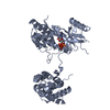
 PDBj
PDBj


 Assembly


 Mass: 294.308 Da / Num. of mol.: 1 / Source method: obtained synthetically / Formula: C16H14N4O2 / Feature type: SUBJECT OF INVESTIGATION
 Mass: 18.015 Da / Num. of mol.: 106 / Source method: isolated from a natural source / Formula: H2O
 Sample preparation
 Processing