Resolution: 1.85→30.975 Å / Cor.coef. Fo:Fc: 0.961 / SU ML: 0.072 / Cross valid method: THROUGHOUT / ESU R: 0.155 / ESU R Free: 0.151 Details: Hydrogens have been added in their riding positions.
Rfactor
Num. reflection
% reflection
Selection details
Rfree
0.21528
800
5.1 %
Random selection
Rwork
0.15986
15608
-
-
all
0.161
-
-
-
obs
0.1607
15608
95.316 %
-
Solvent computation
Ion probe radii: 0.8 Å / Shrinkage radii: 0.8 Å / VDW probe radii: 1.2 Å / Solvent model: MASK BULK SOLVENT
Displacement parameters
Biso mean: 18.205 Å2
Baniso -1
Baniso -2
Baniso -3
1-
0.041 Å2
-0 Å2
-0 Å2
2-
-
-0.524 Å2
0 Å2
3-
-
-
0.482 Å2
Refinement step
Cycle: LAST / Resolution: 1.85→30.975 Å
Protein
Nucleic acid
Ligand
Solvent
Total
Num. atoms
1602
0
47
251
1900
Refine LS restraints
Refine-ID
Type
Dev ideal
Dev ideal target
Number
X-RAY DIFFRACTION
r_bond_refined_d
0.011
0.013
1786
X-RAY DIFFRACTION
r_bond_other_d
0.033
0.017
1750
X-RAY DIFFRACTION
r_angle_refined_deg
1.727
1.649
2451
X-RAY DIFFRACTION
r_angle_other_deg
2.289
1.581
4050
X-RAY DIFFRACTION
r_dihedral_angle_1_deg
5.814
5
237
X-RAY DIFFRACTION
r_dihedral_angle_2_deg
31.949
21.939
98
X-RAY DIFFRACTION
r_dihedral_angle_3_deg
15.557
15
323
X-RAY DIFFRACTION
r_dihedral_angle_4_deg
18.686
15
17
X-RAY DIFFRACTION
r_chiral_restr
0.08
0.2
236
X-RAY DIFFRACTION
r_gen_planes_refined
0.011
0.02
2052
X-RAY DIFFRACTION
r_gen_planes_other
0.016
0.02
384
X-RAY DIFFRACTION
r_nbd_refined
0.208
0.2
385
X-RAY DIFFRACTION
r_symmetry_nbd_other
0.22
0.2
1606
X-RAY DIFFRACTION
r_nbtor_refined
0.166
0.2
854
X-RAY DIFFRACTION
r_symmetry_nbtor_other
0.075
0.2
753
X-RAY DIFFRACTION
r_xyhbond_nbd_refined
0.186
0.2
174
X-RAY DIFFRACTION
r_symmetry_xyhbond_nbd_other
0.139
0.2
1
X-RAY DIFFRACTION
r_symmetry_nbd_refined
0.165
0.2
10
X-RAY DIFFRACTION
r_nbd_other
0.193
0.2
53
X-RAY DIFFRACTION
r_symmetry_xyhbond_nbd_refined
0.222
0.2
31
X-RAY DIFFRACTION
r_mcbond_it
1.676
1.648
865
X-RAY DIFFRACTION
r_mcbond_other
1.676
1.647
864
X-RAY DIFFRACTION
r_mcangle_it
2.528
2.46
1089
X-RAY DIFFRACTION
r_mcangle_other
2.527
2.46
1090
X-RAY DIFFRACTION
r_scbond_it
2.379
1.986
921
X-RAY DIFFRACTION
r_scbond_other
2.376
1.981
919
X-RAY DIFFRACTION
r_scangle_it
3.741
2.863
1348
X-RAY DIFFRACTION
r_scangle_other
3.74
2.865
1349
X-RAY DIFFRACTION
r_lrange_it
5.779
20.617
2117
X-RAY DIFFRACTION
r_lrange_other
5.698
20.165
2071
LS refinement shell
Refine-ID: X-RAY DIFFRACTION
Resolution (Å)
Rfactor Rfree
Num. reflection Rfree
% reflection Rfree (%)
Rfactor Rwork
Num. reflection Rwork
% reflection obs (%)
1.85-1.898
0.262
56
6.9
0.269
751
69.2704
1.898-1.95
0.327
52
5.2
0.235
950
86.9792
1.95-2.006
0.274
42
4
0.202
1004
91.3537
2.006-2.068
0.257
46
4.5
0.194
980
93.4426
2.068-2.136
0.261
46
4.6
0.18
951
95.4981
2.136-2.21
0.268
55
5.4
0.165
964
97.9808
2.21-2.294
0.165
48
4.9
0.151
938
99.2951
2.294-2.387
0.215
51
5.3
0.148
903
100
2.387-2.493
0.178
43
4.6
0.145
882
100
2.493-2.614
0.199
48
5.4
0.152
849
99.8886
2.614-2.755
0.18
41
4.8
0.156
805
100
2.755-2.921
0.182
41
5.1
0.146
759
100
2.921-3.122
0.255
36
4.8
0.153
716
100
3.122-3.371
0.184
48
6.7
0.154
664
99.8597
3.371-3.69
0.204
34
5.2
0.126
619
100
3.69-4.122
0.154
34
5.8
0.119
555
99.6616
4.122-4.753
0.203
31
5.7
0.127
512
99.8162
4.753-5.804
0.233
22
4.8
0.178
440
100
5.804-8.136
0.273
17
4.6
0.201
352
100
8.136-30.975
0.291
9
4
0.199
214
96.5368
+
About Yorodumi
-
News
-
Feb 9, 2022. New format data for meta-information of EMDB entries
New format data for meta-information of EMDB entries
Version 3 of the EMDB header file is now the official format.
The previous official version 1.9 will be removed from the archive.
In the structure databanks used in Yorodumi, some data are registered as the other names, "COVID-19 virus" and "2019-nCoV". Here are the details of the virus and the list of structure data.
Jan 31, 2019. EMDB accession codes are about to change! (news from PDBe EMDB page)
EMDB accession codes are about to change! (news from PDBe EMDB page)
The allocation of 4 digits for EMDB accession codes will soon come to an end. Whilst these codes will remain in use, new EMDB accession codes will include an additional digit and will expand incrementally as the available range of codes is exhausted. The current 4-digit format prefixed with “EMD-” (i.e. EMD-XXXX) will advance to a 5-digit format (i.e. EMD-XXXXX), and so on. It is currently estimated that the 4-digit codes will be depleted around Spring 2019, at which point the 5-digit format will come into force.
The EM Navigator/Yorodumi systems omit the EMD- prefix.
Related info.:Q: What is EMD? / ID/Accession-code notation in Yorodumi/EM Navigator
Yorodumi is a browser for structure data from EMDB, PDB, SASBDB, etc.
This page is also the successor to EM Navigator detail page, and also detail information page/front-end page for Omokage search.
The word "yorodu" (or yorozu) is an old Japanese word meaning "ten thousand". "mi" (miru) is to see.
Related info.:EMDB / PDB / SASBDB / Comparison of 3 databanks / Yorodumi Search / Aug 31, 2016. New EM Navigator & Yorodumi / Yorodumi Papers / Jmol/JSmol / Function and homology information / Changes in new EM Navigator and Yorodumi
 Movie
Movie Controller

 Yorodumi
 Open data
 Basic information
 Components
 Keywords
 Function and homology information
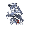
 Erythrobacter litoralis HTCC2594 (bacteria)
 X-RAY DIFFRACTION /
  Authors
 Czech Republic, European Union, 3items
  Citation
 Structure visualization
 Downloads & links
 Other downloads
 PDBj
PDBj


 Assembly
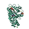


 Mass: 456.344 Da / Num. of mol.: 1 / Source method: obtained synthetically / Formula: C17H21N4O9P / Feature type: SUBJECT OF INVESTIGATION
 Mass: 35.453 Da / Num. of mol.: 2 / Source method: obtained synthetically / Formula: Cl
 Mass: 24.305 Da / Num. of mol.: 2 / Source method: obtained synthetically / Formula: Mg
 Mass: 195.237 Da / Num. of mol.: 1 / Source method: obtained synthetically / Formula: C6H13NO4S / Comment: pH buffer*YM
 Sample preparation
 Processing