Resolution: 2.918→56.55 Å / Cor.coef. Fo:Fc: 0.94 / Cor.coef. Fo:Fc free: 0.861 / SU B: 23.235 / SU ML: 0.404 / Cross valid method: FREE R-VALUE / ESU R Free: 0.443 Details: Hydrogens have been added in their riding positions
Rfactor
Num. reflection
% reflection
Rfree
0.2746
524
5.125 %
Rwork
0.1931
9700
-
all
0.197
-
-
obs
-
10224
99.873 %
Solvent computation
Ion probe radii: 0.8 Å / Shrinkage radii: 0.8 Å / VDW probe radii: 1.2 Å / Solvent model: MASK BULK SOLVENT
Displacement parameters
Biso mean: 54.013 Å2
Baniso -1
Baniso -2
Baniso -3
1-
-3.679 Å2
-0 Å2
-2.008 Å2
2-
-
1.848 Å2
0 Å2
3-
-
-
2.491 Å2
Refinement step
Cycle: LAST / Resolution: 2.918→56.55 Å
Protein
Nucleic acid
Ligand
Solvent
Total
Num. atoms
2809
0
15
24
2848
Refine LS restraints
Refine-ID
Type
Dev ideal
Dev ideal target
Number
X-RAY DIFFRACTION
r_bond_refined_d
0.006
0.013
2902
X-RAY DIFFRACTION
r_bond_other_d
0.001
0.014
2668
X-RAY DIFFRACTION
r_angle_refined_deg
1.537
1.644
3947
X-RAY DIFFRACTION
r_angle_other_deg
1.166
1.576
6114
X-RAY DIFFRACTION
r_dihedral_angle_1_deg
8.98
5
352
X-RAY DIFFRACTION
r_dihedral_angle_2_deg
30.156
19.885
174
X-RAY DIFFRACTION
r_dihedral_angle_3_deg
19.318
15
452
X-RAY DIFFRACTION
r_dihedral_angle_4_deg
20.859
15
32
X-RAY DIFFRACTION
r_chiral_restr
0.055
0.2
348
X-RAY DIFFRACTION
r_gen_planes_refined
0.006
0.02
3305
X-RAY DIFFRACTION
r_gen_planes_other
0.002
0.02
743
X-RAY DIFFRACTION
r_nbd_refined
0.185
0.2
524
X-RAY DIFFRACTION
r_symmetry_nbd_other
0.197
0.2
2605
X-RAY DIFFRACTION
r_nbtor_refined
0.165
0.2
1302
X-RAY DIFFRACTION
r_symmetry_nbtor_other
0.078
0.2
1434
X-RAY DIFFRACTION
r_xyhbond_nbd_refined
0.144
0.2
75
X-RAY DIFFRACTION
r_symmetry_nbd_refined
0.211
0.2
7
X-RAY DIFFRACTION
r_nbd_other
0.236
0.2
44
X-RAY DIFFRACTION
r_symmetry_xyhbond_nbd_refined
0.055
0.2
2
X-RAY DIFFRACTION
r_mcbond_it
3.661
5.504
1408
X-RAY DIFFRACTION
r_mcbond_other
3.661
5.504
1407
X-RAY DIFFRACTION
r_mcangle_it
5.992
8.242
1754
X-RAY DIFFRACTION
r_mcangle_other
5.991
8.242
1755
X-RAY DIFFRACTION
r_scbond_it
4.127
6.04
1494
X-RAY DIFFRACTION
r_scbond_other
4.077
6.014
1482
X-RAY DIFFRACTION
r_scangle_it
6.571
8.88
2191
X-RAY DIFFRACTION
r_scangle_other
6.547
8.843
2173
X-RAY DIFFRACTION
r_lrange_it
9.702
62.115
3027
X-RAY DIFFRACTION
r_lrange_other
9.701
62.118
3028
LS refinement shell
Resolution (Å)
Rfactor Rfree
Num. reflection Rfree
Rfactor Rwork
Num. reflection Rwork
Refine-ID
% reflection obs (%)
2.918-2.994
0.357
43
0.31
675
X-RAY DIFFRACTION
99.1713
2.994-3.076
0.336
37
0.308
694
X-RAY DIFFRACTION
99.5913
3.076-3.165
0.376
39
0.293
689
X-RAY DIFFRACTION
100
3.165-3.262
0.264
34
0.269
644
X-RAY DIFFRACTION
99.8527
3.262-3.369
0.298
39
0.24
649
X-RAY DIFFRACTION
99.8549
3.369-3.488
0.305
32
0.219
620
X-RAY DIFFRACTION
100
3.488-3.619
0.224
20
0.189
599
X-RAY DIFFRACTION
100
3.619-3.767
0.297
36
0.219
593
X-RAY DIFFRACTION
100
3.767-3.934
0.333
24
0.2
532
X-RAY DIFFRACTION
100
3.934-4.126
0.33
26
0.193
546
X-RAY DIFFRACTION
100
4.126-4.349
0.216
29
0.171
499
X-RAY DIFFRACTION
100
4.349-4.612
0.291
24
0.135
480
X-RAY DIFFRACTION
99.802
4.612-4.93
0.146
18
0.129
453
X-RAY DIFFRACTION
100
4.93-5.324
0.246
23
0.124
416
X-RAY DIFFRACTION
100
5.324-5.831
0.216
24
0.156
382
X-RAY DIFFRACTION
100
5.831-6.517
0.301
24
0.16
334
X-RAY DIFFRACTION
99.7215
6.517-7.521
0.327
13
0.182
320
X-RAY DIFFRACTION
100
7.521-9.201
0.259
18
0.162
252
X-RAY DIFFRACTION
100
9.201-12.969
0.19
17
0.135
204
X-RAY DIFFRACTION
100
+
About Yorodumi
-
News
-
Feb 9, 2022. New format data for meta-information of EMDB entries
New format data for meta-information of EMDB entries
Version 3 of the EMDB header file is now the official format.
The previous official version 1.9 will be removed from the archive.
In the structure databanks used in Yorodumi, some data are registered as the other names, "COVID-19 virus" and "2019-nCoV". Here are the details of the virus and the list of structure data.
Jan 31, 2019. EMDB accession codes are about to change! (news from PDBe EMDB page)
EMDB accession codes are about to change! (news from PDBe EMDB page)
The allocation of 4 digits for EMDB accession codes will soon come to an end. Whilst these codes will remain in use, new EMDB accession codes will include an additional digit and will expand incrementally as the available range of codes is exhausted. The current 4-digit format prefixed with “EMD-” (i.e. EMD-XXXX) will advance to a 5-digit format (i.e. EMD-XXXXX), and so on. It is currently estimated that the 4-digit codes will be depleted around Spring 2019, at which point the 5-digit format will come into force.
The EM Navigator/Yorodumi systems omit the EMD- prefix.
Related info.:Q: What is EMD? / ID/Accession-code notation in Yorodumi/EM Navigator
Yorodumi is a browser for structure data from EMDB, PDB, SASBDB, etc.
This page is also the successor to EM Navigator detail page, and also detail information page/front-end page for Omokage search.
The word "yorodu" (or yorozu) is an old Japanese word meaning "ten thousand". "mi" (miru) is to see.
Related info.:EMDB / PDB / SASBDB / Comparison of 3 databanks / Yorodumi Search / Aug 31, 2016. New EM Navigator & Yorodumi / Yorodumi Papers / Jmol/JSmol / Function and homology information / Changes in new EM Navigator and Yorodumi
 Movie
Movie Controller


 Open data
 Basic information
 Components
 Keywords
 Function and homology information
 Homo sapiens (human)
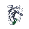
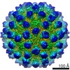 Murine norovirus 1
 X-RAY DIFFRACTION /
  Authors
 United Kingdom, 1items
  Citation
 Structure visualization
 Downloads & links
 Other downloads


 PDBj
PDBj

 Assembly
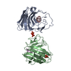


 Mass: 94.971 Da / Num. of mol.: 3 / Source method: obtained synthetically / Formula: PO4
 Mass: 18.015 Da / Num. of mol.: 24 / Source method: isolated from a natural source / Formula: H2O
 Sample preparation
 Processing