 Movie
Movie Controller


+ Open data

- Basic information
| Entry | Database: PDB / ID: 8a3x | ||||||
|---|---|---|---|---|---|---|---|
| Title | Imine Reductase from Ensifer adhaerens in complex with NADP+ | ||||||
 Components Components | NAD_binding_2 domain-containing protein | ||||||
 Keywords Keywords | OXIDOREDUCTASE / Imine Reductase / IRED / NADP+ / amine | ||||||
| Function / homology |  Function and homology information Function and homology information | ||||||
| Biological species |  Ensifer adhaerens (bacteria) Ensifer adhaerens (bacteria) | ||||||
| Method |  X-RAY DIFFRACTION / SYNCHROTRON / MOLECULAR REPLACEMENT / Resolution: 2.58 Å X-RAY DIFFRACTION / SYNCHROTRON / MOLECULAR REPLACEMENT / Resolution: 2.58 Å | ||||||
 Authors Authors | Gilio, A.K. / Grogan, G. | ||||||
| Funding support | 1items
| ||||||
 Citation Citation | Journal: Acs Catalysis / Year: 2023 Title: A Reductive Aminase Switches to Imine Reductase Mode for a Bulky Amine Substrate. Authors: Gilio, A.K. / Thorpe, T.W. / Heyam, A. / Petchey, M.R. / Pogranyi, B. / France, S.P. / Howard, R.M. / Karmilowicz, M.J. / Lewis, R. / Turner, N. / Grogan, G. | ||||||
| History |
|
- Structure visualization
| Structure viewer | Molecule: MolmilJmol/JSmol |
|---|
- Downloads & links
-Download
| PDBx/mmCIF format | 8a3x.cif.gz | 225.7 KB | Display | PDBx/mmCIF format |
|---|---|---|---|---|
| PDB format | pdb8a3x.ent.gz | 181.3 KB | Display | PDB format |
| PDBx/mmJSON format | 8a3x.json.gz | Tree view | PDBx/mmJSON format | |
| Others |  Other downloads Other downloads |
-Validation report
| Summary document | 8a3x_validation.pdf.gz | 1.6 MB | Display | wwPDB validaton report |
|---|---|---|---|---|
| Full document | 8a3x_full_validation.pdf.gz | 1.7 MB | Display | |
| Data in XML | 8a3x_validation.xml.gz | 43 KB | Display | |
| Data in CIF | 8a3x_validation.cif.gz | 57.6 KB | Display | |
| Arichive directory | https://data.pdbj.org/pub/pdb/validation_reports/a3/8a3xftp://data.pdbj.org/pub/pdb/validation_reports/a3/8a3x | HTTPS FTP |
-Related structure data
| Related structure data |  8a5zC  5ocmS S: Starting model for refinement C: citing same article ( |
|---|---|
| Similar structure data |
-Links
 PDBj
PDBj
- Assembly
| Deposited unit | 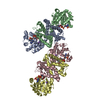
| ||||||||||||||||||||||||||||||||||||||||||||||||||||||||||||||||||||||||||||||||||||||||||||||||||||||||||||||||||||||||||||||||||||||||||||||||||||||||||||||||||||||||||||||||
|---|---|---|---|---|---|---|---|---|---|---|---|---|---|---|---|---|---|---|---|---|---|---|---|---|---|---|---|---|---|---|---|---|---|---|---|---|---|---|---|---|---|---|---|---|---|---|---|---|---|---|---|---|---|---|---|---|---|---|---|---|---|---|---|---|---|---|---|---|---|---|---|---|---|---|---|---|---|---|---|---|---|---|---|---|---|---|---|---|---|---|---|---|---|---|---|---|---|---|---|---|---|---|---|---|---|---|---|---|---|---|---|---|---|---|---|---|---|---|---|---|---|---|---|---|---|---|---|---|---|---|---|---|---|---|---|---|---|---|---|---|---|---|---|---|---|---|---|---|---|---|---|---|---|---|---|---|---|---|---|---|---|---|---|---|---|---|---|---|---|---|---|---|---|---|---|---|---|
| 1 | 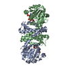
| ||||||||||||||||||||||||||||||||||||||||||||||||||||||||||||||||||||||||||||||||||||||||||||||||||||||||||||||||||||||||||||||||||||||||||||||||||||||||||||||||||||||||||||||||
| 2 | 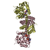
| ||||||||||||||||||||||||||||||||||||||||||||||||||||||||||||||||||||||||||||||||||||||||||||||||||||||||||||||||||||||||||||||||||||||||||||||||||||||||||||||||||||||||||||||||
| Unit cell |
| ||||||||||||||||||||||||||||||||||||||||||||||||||||||||||||||||||||||||||||||||||||||||||||||||||||||||||||||||||||||||||||||||||||||||||||||||||||||||||||||||||||||||||||||||
| Noncrystallographic symmetry (NCS) | NCS domain:
NCS domain segments: Component-ID: _ / Refine code: _
NCS ensembles :
|
-Components
| #1: Protein | Mass: 31105.219 Da / Num. of mol.: 4 Source method: isolated from a genetically manipulated source Source: (gene. exp.) Ensifer adhaerens (bacteria) / Gene: AC244_29830 / Plasmid: pET28a / Production host: #2: Chemical | ChemComp-NAP /   Mass: 743.405 Da / Num. of mol.: 4 / Source method: obtained synthetically / Formula: C21H28N7O17P3 / Feature type: SUBJECT OF INVESTIGATION Mass: 743.405 Da / Num. of mol.: 4 / Source method: obtained synthetically / Formula: C21H28N7O17P3 / Feature type: SUBJECT OF INVESTIGATION#3: Chemical | ChemComp-K /   Mass: 39.098 Da / Num. of mol.: 4 / Source method: obtained synthetically / Formula: K Mass: 39.098 Da / Num. of mol.: 4 / Source method: obtained synthetically / Formula: K#4: Water | ChemComp-HOH / |  Mass: 18.015 Da / Num. of mol.: 36 / Source method: isolated from a natural source / Formula: H2O Mass: 18.015 Da / Num. of mol.: 36 / Source method: isolated from a natural source / Formula: H2OHas ligand of interest | Y | |
|---|
-Experimental details
-Experiment
| Experiment | Method: X-RAY DIFFRACTION / Number of used crystals: 1 |
|---|
- Sample preparation
| Crystal | Density Matthews: 2.44 Å3/Da / Density % sol: 49.58 % |
|---|---|
| Crystal grow | Temperature: 279 K / Method: vapor diffusion, sitting drop / pH: 7.1 Details: 30% (w/v) PEG 2000 MME; 0.2 M KSCN; protein at 50 mg mL-1 Temp details: Grown at 6 deg C |
-Data collection
| Diffraction | Mean temperature: 120 K / Serial crystal experiment: N |
|---|---|
| Diffraction source | Source: SYNCHROTRON / Site: Diamond  / Beamline: I04 / Wavelength: 0.9795 Å / Beamline: I04 / Wavelength: 0.9795 Å |
| Detector | Type: DECTRIS EIGER2 XE 16M / Detector: PIXEL / Date: Feb 20, 2020 |
| Radiation | Protocol: SINGLE WAVELENGTH / Monochromatic (M) / Laue (L): M / Scattering type: x-ray |
| Radiation wavelength | Wavelength: 0.9795 Å / Relative weight: 1 |
| Reflection | Resolution: 2.58→58 Å / Num. obs: 38177 / % possible obs: 99.7 % / Redundancy: 4.1 % / Biso Wilson estimate: 31 Å2 / CC1/2: 0.99 / Rmerge(I) obs: 0.17 / Rpim(I) all: 0.15 / Net I/σ(I): 4.4 |
| Reflection shell | Resolution: 2.58→2.69 Å / Rmerge(I) obs: 1.17 / Mean I/σ(I) obs: 0.9 / Num. unique obs: 4621 / CC1/2: 0.67 / Rpim(I) all: 0.97 |
- Processing
| Software |
| |||||||||||||||||||||||||||||||||||||||||||||||||||||||||||||||||
|---|---|---|---|---|---|---|---|---|---|---|---|---|---|---|---|---|---|---|---|---|---|---|---|---|---|---|---|---|---|---|---|---|---|---|---|---|---|---|---|---|---|---|---|---|---|---|---|---|---|---|---|---|---|---|---|---|---|---|---|---|---|---|---|---|---|---|
| Refinement | Method to determine structure: MOLECULAR REPLACEMENT Starting model: 5OCM Resolution: 2.58→58 Å / Cor.coef. Fo:Fc: 0.931 / Cor.coef. Fo:Fc free: 0.905 / SU B: 24.897 / SU ML: 0.452 / Cross valid method: THROUGHOUT / σ(F): 0 / ESU R: 1.714 / ESU R Free: 0.363 / Stereochemistry target values: MAXIMUM LIKELIHOOD Details: HYDROGENS HAVE BEEN ADDED IN THE RIDING POSITIONS U VALUES : REFINED INDIVIDUALLY
| |||||||||||||||||||||||||||||||||||||||||||||||||||||||||||||||||
| Solvent computation | Ion probe radii: 0.8 Å / Shrinkage radii: 0.8 Å / VDW probe radii: 1.2 Å / Solvent model: MASK | |||||||||||||||||||||||||||||||||||||||||||||||||||||||||||||||||
| Displacement parameters | Biso max: 118.07 Å2 / Biso mean: 52.967 Å2 / Biso min: 16.7 Å2
| |||||||||||||||||||||||||||||||||||||||||||||||||||||||||||||||||
| Refinement step | Cycle: final / Resolution: 2.58→58 Å
| |||||||||||||||||||||||||||||||||||||||||||||||||||||||||||||||||
| Refine LS restraints |
| |||||||||||||||||||||||||||||||||||||||||||||||||||||||||||||||||
| Refine LS restraints NCS | Refine-ID: X-RAY DIFFRACTION / Type: interatomic distance / Weight position: 0.05
| |||||||||||||||||||||||||||||||||||||||||||||||||||||||||||||||||
| LS refinement shell | Resolution: 2.58→2.647 Å / Rfactor Rfree error: 0 / Total num. of bins used: 20
|
