 Movie
Movie Controller

[English] 日本語
 Yorodumi
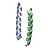
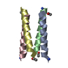
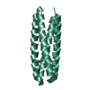
- PDB-8a3g: X-ray crystal structure of a de novo designed antiparallel coiled... -
+ Open data

- Basic information
| Entry | Database: PDB / ID: 8a3g | |||||||||||||||||||||||||||||||||||||
|---|---|---|---|---|---|---|---|---|---|---|---|---|---|---|---|---|---|---|---|---|---|---|---|---|---|---|---|---|---|---|---|---|---|---|---|---|---|---|
| Title | X-ray crystal structure of a de novo designed antiparallel coiled-coil homotetramer with 4 heptad repeats, apCC-Tet* | |||||||||||||||||||||||||||||||||||||
 Components Components | apCC-Tet* Keywords KeywordsDE NOVO PROTEIN / coiled coil / 4-helix bundle / de novo protein design / peptide assembly | Function / homology | ACETATE ION |  Function and homology information Function and homology informationBiological species | synthetic construct (others) | Method |  X-RAY DIFFRACTION / SYNCHROTRON / AB INITIO PHASING / Resolution: 0.96 Å X-RAY DIFFRACTION / SYNCHROTRON / AB INITIO PHASING / Resolution: 0.96 Å  Authors AuthorsNaudin, E.A. / Mylemans, B. / Albanese, K.I. / Woolfson, D.N. | Funding support | |  United Kingdom, 4items United Kingdom, 4items
 CitationJournal: Chem Sci / Year: 2022 CitationJournal: Chem Sci / Year: 2022Title: From peptides to proteins: coiled-coil tetramers to single-chain 4-helix bundles. Authors: Naudin, E.A. / Albanese, K.I. / Smith, A.J. / Mylemans, B. / Baker, E.G. / Weiner, O.D. / Andrews, D.M. / Tigue, N. / Savery, N.J. / Woolfson, D.N. History |
|
- Structure visualization
| Structure viewer | Molecule: MolmilJmol/JSmol |
|---|
- Downloads & links
-Download
| PDBx/mmCIF format | 8a3g.cif.gz | 39.2 KB | Display | PDBx/mmCIF format |
|---|---|---|---|---|
| PDB format | pdb8a3g.ent.gz | 26.9 KB | Display | PDB format |
| PDBx/mmJSON format | 8a3g.json.gz | Tree view | PDBx/mmJSON format | |
| Others |  Other downloads Other downloads |
-Validation report
| Arichive directory | https://data.pdbj.org/pub/pdb/validation_reports/a3/8a3gftp://data.pdbj.org/pub/pdb/validation_reports/a3/8a3g | HTTPS FTP |
|---|
-Related structure data
-Links
 PDBj
PDBj
- Assembly
| Deposited unit | 
| ||||||||||
|---|---|---|---|---|---|---|---|---|---|---|---|
| 1 | 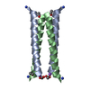
| ||||||||||
| Unit cell |
|
-Components
| #1: Protein/peptide | Mass: 3463.076 Da / Num. of mol.: 2 / Source method: obtained synthetically / Source: (synth.) synthetic construct (others) #2: Chemical | ChemComp-ACT / |   Mass: 59.044 Da / Num. of mol.: 1 / Source method: obtained synthetically / Formula: C2H3O2 Mass: 59.044 Da / Num. of mol.: 1 / Source method: obtained synthetically / Formula: C2H3O2#3: Chemical | ChemComp-NA / |   Mass: 22.990 Da / Num. of mol.: 1 / Source method: obtained synthetically / Formula: Na Mass: 22.990 Da / Num. of mol.: 1 / Source method: obtained synthetically / Formula: Na#4: Water | ChemComp-HOH / |  Mass: 18.015 Da / Num. of mol.: 39 / Source method: isolated from a natural source / Formula: H2O Mass: 18.015 Da / Num. of mol.: 39 / Source method: isolated from a natural source / Formula: H2OHas ligand of interest | N | Has protein modification | Y | |
|---|
-Experimental details
-Experiment
| Experiment | Method: X-RAY DIFFRACTION / Number of used crystals: 1 |
|---|
- Sample preparation
| Crystal | Density Matthews: 1.84 Å3/Da / Density % sol: 33.17 % |
|---|---|
| Crystal grow | Temperature: 293 K / Method: vapor diffusion, sitting drop / pH: 5 Details: 2.0 M Ammonium sulfate, 0.1 M Sodium acetate, pH 5.0 |
-Data collection
| Diffraction | Mean temperature: 100 K / Serial crystal experiment: N | ||||||||||||||||||||||||||||||
|---|---|---|---|---|---|---|---|---|---|---|---|---|---|---|---|---|---|---|---|---|---|---|---|---|---|---|---|---|---|---|---|
| Diffraction source | Source: SYNCHROTRON / Site: Diamond / Beamline: I24 / Wavelength: 0.85 Å | ||||||||||||||||||||||||||||||
| Detector | Type: DECTRIS PILATUS 6M / Detector: PIXEL / Date: Jan 29, 2021 | ||||||||||||||||||||||||||||||
| Radiation | Protocol: SINGLE WAVELENGTH / Monochromatic (M) / Laue (L): M / Scattering type: x-ray | ||||||||||||||||||||||||||||||
| Radiation wavelength | Wavelength: 0.85 Å / Relative weight: 1 | ||||||||||||||||||||||||||||||
| Reflection | Resolution: 0.96→43.77 Å / Num. obs: 31082 / % possible obs: 96.1 % / Redundancy: 20.8 % / Biso Wilson estimate: 8.99 Å2 / CC1/2: 1 / Rmerge(I) obs: 0.042 / Rpim(I) all: 0.009 / Rrim(I) all: 0.043 / Net I/σ(I): 30.3 / Num. measured all: 645142 / Scaling rejects: 144 | ||||||||||||||||||||||||||||||
| Reflection shell | Diffraction-ID: 1
|
- Processing
| Software |
| ||||||||||||||||||||||||||||||||||||||||||||||||||||||||||||||||||||||||||||||||||||
|---|---|---|---|---|---|---|---|---|---|---|---|---|---|---|---|---|---|---|---|---|---|---|---|---|---|---|---|---|---|---|---|---|---|---|---|---|---|---|---|---|---|---|---|---|---|---|---|---|---|---|---|---|---|---|---|---|---|---|---|---|---|---|---|---|---|---|---|---|---|---|---|---|---|---|---|---|---|---|---|---|---|---|---|---|---|
| Refinement | Method to determine structure: AB INITIO PHASING / Resolution: 0.96→26.86 Å / SU ML: 0.05 / Cross valid method: THROUGHOUT / σ(F): 1.97 / Phase error: 15.91 / Stereochemistry target values: ML
| ||||||||||||||||||||||||||||||||||||||||||||||||||||||||||||||||||||||||||||||||||||
| Solvent computation | Shrinkage radii: 0.9 Å / VDW probe radii: 1.11 Å / Solvent model: FLAT BULK SOLVENT MODEL | ||||||||||||||||||||||||||||||||||||||||||||||||||||||||||||||||||||||||||||||||||||
| Displacement parameters | Biso max: 39.5 Å2 / Biso mean: 12.7871 Å2 / Biso min: 5.99 Å2 | ||||||||||||||||||||||||||||||||||||||||||||||||||||||||||||||||||||||||||||||||||||
| Refinement step | Cycle: final / Resolution: 0.96→26.86 Å
| ||||||||||||||||||||||||||||||||||||||||||||||||||||||||||||||||||||||||||||||||||||
| LS refinement shell | Refine-ID: X-RAY DIFFRACTION / Rfactor Rfree error: 0 / Total num. of bins used: 11
|
