- PDB-8a2a: EGFR kinase domain in complex with 2-(6,7-dihydro-5H-pyrrolo[1,2-... -
+
Open data
ID or keywords:
Loading...
-
Basic information
Entry
Database: PDB / ID: 8a2a
Title
EGFR kinase domain in complex with 2-(6,7-dihydro-5H-pyrrolo[1,2-c]imidazol-1-yl)-2-[6-[2-[4-[[4-(hydroxymethyl)-1-piperidyl]methyl]phenyl]ethynyl]-1-oxo-4-(trifluoromethyl)isoindolin-2-yl]-N-thiazol-2-yl-acetamide (form 2)
Components
Epidermal growth factor receptor
Keywords
TRANSFERASE / KINASE INHIBITOR
Function / homology
Function and homology information
multivesicular body, internal vesicle lumen / negative regulation of cardiocyte differentiation / Shc-EGFR complex / positive regulation of protein kinase C signaling / Inhibition of Signaling by Overexpressed EGFR / epidermal growth factor receptor activity / EGFR interacts with phospholipase C-gamma / regulation of peptidyl-tyrosine phosphorylation / epidermal growth factor binding / response to UV-A ...multivesicular body, internal vesicle lumen / negative regulation of cardiocyte differentiation / Shc-EGFR complex / positive regulation of protein kinase C signaling / Inhibition of Signaling by Overexpressed EGFR / epidermal growth factor receptor activity / EGFR interacts with phospholipase C-gamma / regulation of peptidyl-tyrosine phosphorylation / epidermal growth factor binding / response to UV-A / PLCG1 events in ERBB2 signaling / ERBB2-EGFR signaling pathway / morphogenesis of an epithelial fold / PTK6 promotes HIF1A stabilization / ERBB2 Activates PTK6 Signaling / digestive tract morphogenesis / Signaling by EGFR / intracellular vesicle / negative regulation of epidermal growth factor receptor signaling pathway / eyelid development in camera-type eye / cerebral cortex cell migration / protein insertion into membrane / ERBB2 Regulates Cell Motility / protein tyrosine kinase activator activity / Respiratory syncytial virus (RSV) attachment and entry / Signaling by ERBB4 / PI3K events in ERBB2 signaling / positive regulation of phosphorylation / positive regulation of peptidyl-serine phosphorylation / Estrogen-dependent nuclear events downstream of ESR-membrane signaling / hair follicle development / MAP kinase kinase kinase activity / GAB1 signalosome / positive regulation of G1/S transition of mitotic cell cycle / embryonic placenta development / salivary gland morphogenesis / Signaling by ERBB2 / TFAP2 (AP-2) family regulates transcription of growth factors and their receptors / GRB2 events in EGFR signaling / transmembrane receptor protein tyrosine kinase activity / SHC1 events in EGFR signaling / EGFR Transactivation by Gastrin / GRB2 events in ERBB2 signaling / SHC1 events in ERBB2 signaling / ossification / basal plasma membrane / cellular response to epidermal growth factor stimulus / positive regulation of DNA repair / positive regulation of DNA replication / epithelial cell proliferation / Signal transduction by L1 / positive regulation of epithelial cell proliferation / positive regulation of protein localization to plasma membrane / cellular response to amino acid stimulus / NOTCH3 Activation and Transmission of Signal to the Nucleus / phosphatidylinositol 3-kinase/protein kinase B signal transduction / cellular response to estradiol stimulus / clathrin-coated endocytic vesicle membrane / EGFR downregulation / Signaling by ERBB2 TMD/JMD mutants / cell-cell adhesion / Constitutive Signaling by EGFRvIII / receptor protein-tyrosine kinase / Signaling by ERBB2 ECD mutants / Signaling by ERBB2 KD Mutants / negative regulation of protein catabolic process / positive regulation of miRNA transcription / kinase binding / epidermal growth factor receptor signaling pathway / ruffle membrane / Downregulation of ERBB2 signaling / positive regulation of fibroblast proliferation / cell morphogenesis / neuron differentiation / positive regulation of protein phosphorylation / HCMV Early Events / Constitutive Signaling by Aberrant PI3K in Cancer / actin filament binding / cell junction / transmembrane signaling receptor activity / positive regulation of canonical Wnt signaling pathway / PIP3 activates AKT signaling / Cargo recognition for clathrin-mediated endocytosis / Constitutive Signaling by Ligand-Responsive EGFR Cancer Variants / Clathrin-mediated endocytosis / ATPase binding / PI5P, PP2A and IER3 Regulate PI3K/AKT Signaling / virus receptor activity / RAF/MAP kinase cascade / positive regulation of cell growth / double-stranded DNA binding / protein tyrosine kinase activity / early endosome membrane / protein phosphatase binding / nuclear membrane / basolateral plasma membrane / learning or memory / cell surface receptor signaling pathway / Extra-nuclear estrogen signaling / positive regulation of ERK1 and ERK2 cascade Similarity search - Function
: / Epidermal growth factor receptor transmembrane-juxtamembrane segment / Tyrosine protein kinase, EGF/ERB/XmrK receptor / Growth factor receptor domain 4 / Growth factor receptor domain IV / Receptor L-domain / Furin-like cysteine-rich domain / Receptor L-domain superfamily / Furin-like cysteine rich region / Receptor L domain ...: / Epidermal growth factor receptor transmembrane-juxtamembrane segment / Tyrosine protein kinase, EGF/ERB/XmrK receptor / Growth factor receptor domain 4 / Growth factor receptor domain IV / Receptor L-domain / Furin-like cysteine-rich domain / Receptor L-domain superfamily / Furin-like cysteine rich region / Receptor L domain / Furin-like repeat / Furin-like repeats / Growth factor receptor cysteine-rich domain superfamily / : / Tyrosine-protein kinase, catalytic domain / Tyrosine kinase, catalytic domain / Tyrosine protein kinases specific active-site signature. / Tyrosine-protein kinase, active site / Serine-threonine/tyrosine-protein kinase, catalytic domain / Protein tyrosine and serine/threonine kinase / Transferase(Phosphotransferase) domain 1 / Transferase(Phosphotransferase); domain 1 / Protein kinase, ATP binding site / Protein kinases ATP-binding region signature. / Protein kinase domain profile. / Protein kinase domain / Protein kinase-like domain superfamily / Orthogonal Bundle / Mainly Alpha Similarity search - Domain/homology
Resolution: 1.43→57.31 Å / Cor.coef. Fo:Fc: 0.962 / Cor.coef. Fo:Fc free: 0.952 / SU B: 3.066 / SU ML: 0.058 / Cross valid method: THROUGHOUT / σ(F): 0 / ESU R: 0.094 / ESU R Free: 0.096 / Stereochemistry target values: MAXIMUM LIKELIHOOD Details: HYDROGENS HAVE BEEN ADDED IN THE RIDING POSITIONS U VALUES : WITH TLS ADDED
Rfactor
Num. reflection
% reflection
Selection details
Rfree
0.2233
2074
4.8 %
RANDOM
Rwork
0.188
-
-
-
obs
0.1897
40957
67.97 %
-
Solvent computation
Ion probe radii: 0.8 Å / Shrinkage radii: 0.8 Å / VDW probe radii: 1.2 Å / Solvent model: MASK
In the structure databanks used in Yorodumi, some data are registered as the other names, "COVID-19 virus" and "2019-nCoV". Here are the details of the virus and the list of structure data.
Jan 31, 2019. EMDB accession codes are about to change! (news from PDBe EMDB page)
EMDB accession codes are about to change! (news from PDBe EMDB page)
The allocation of 4 digits for EMDB accession codes will soon come to an end. Whilst these codes will remain in use, new EMDB accession codes will include an additional digit and will expand incrementally as the available range of codes is exhausted. The current 4-digit format prefixed with “EMD-” (i.e. EMD-XXXX) will advance to a 5-digit format (i.e. EMD-XXXXX), and so on. It is currently estimated that the 4-digit codes will be depleted around Spring 2019, at which point the 5-digit format will come into force.
The EM Navigator/Yorodumi systems omit the EMD- prefix.
Related info.:Q: What is EMD? / ID/Accession-code notation in Yorodumi/EM Navigator
Yorodumi is a browser for structure data from EMDB, PDB, SASBDB, etc.
This page is also the successor to EM Navigator detail page, and also detail information page/front-end page for Omokage search.
The word "yorodu" (or yorozu) is an old Japanese word meaning "ten thousand". "mi" (miru) is to see.
Related info.:EMDB / PDB / SASBDB / Comparison of 3 databanks / Yorodumi Search / Aug 31, 2016. New EM Navigator & Yorodumi / Yorodumi Papers / Jmol/JSmol / Function and homology information / Changes in new EM Navigator and Yorodumi
 Movie
Movie Controller

 Yorodumi
 Open data
 Basic information
 Components
 Keywords
 Function and homology information
 Homo sapiens (human)
 X-RAY DIFFRACTION /
  Authors
 Switzerland, 1items
  Citation
 Structure visualization
 Downloads & links
 Other downloads


 PDBj
PDBj


 Assembly
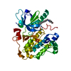

 Spodoptera frugiperda (fall armyworm)
 Mass: 674.735 Da / Num. of mol.: 1 / Source method: obtained synthetically / Formula: C35H33F3N6O3S / Feature type: SUBJECT OF INVESTIGATION
 Mass: 96.063 Da / Num. of mol.: 1 / Source method: obtained synthetically / Formula: SO4
 Mass: 18.015 Da / Num. of mol.: 221 / Source method: isolated from a natural source / Formula: H2O
 Sample preparation
 Processing