 Movie
Movie Controller


+ Open data

- Basic information
| Entry | Database: PDB / ID: 8a1c | |||||||||
|---|---|---|---|---|---|---|---|---|---|---|
| Title | TraI trans-esterase domain from pKM101 (DNA bound) | |||||||||
 Components Components |
| |||||||||
 Keywords Keywords | DNA BINDING PROTEIN / Relaxase / Trans-esterase | |||||||||
| Function / homology |  Function and homology information Function and homology information | |||||||||
| Biological species |  synthetic construct (others) | |||||||||
| Method |  X-RAY DIFFRACTION / SYNCHROTRON / MOLECULAR REPLACEMENT / Resolution: 2.1 Å X-RAY DIFFRACTION / SYNCHROTRON / MOLECULAR REPLACEMENT / Resolution: 2.1 Å | |||||||||
 Authors Authors | Breidenstein, A. / Berntsson, R.P.-A. | |||||||||
| Funding support |  Sweden, 2items Sweden, 2items
| |||||||||
 Citation Citation | Journal: Life Sci Alliance / Year: 2023 Title: Structural and functional characterization of TraI from pKM101 reveals basis for DNA processing. Authors: Breidenstein, A. / Ter Beek, J. / Berntsson, R.P. | |||||||||
| History |
|
- Structure visualization
| Structure viewer | Molecule: MolmilJmol/JSmol |
|---|
- Downloads & links
-Download
| PDBx/mmCIF format | 8a1c.cif.gz | 201.2 KB | Display | PDBx/mmCIF format |
|---|---|---|---|---|
| PDB format | pdb8a1c.ent.gz | 160.2 KB | Display | PDB format |
| PDBx/mmJSON format | 8a1c.json.gz | Tree view | PDBx/mmJSON format | |
| Others |  Other downloads Other downloads |
-Validation report
| Summary document | 8a1c_validation.pdf.gz | 560.6 KB | Display | wwPDB validaton report |
|---|---|---|---|---|
| Full document | 8a1c_full_validation.pdf.gz | 564.3 KB | Display | |
| Data in XML | 8a1c_validation.xml.gz | 12.7 KB | Display | |
| Data in CIF | 8a1c_validation.cif.gz | 16.7 KB | Display | |
| Arichive directory | https://data.pdbj.org/pub/pdb/validation_reports/a1/8a1cftp://data.pdbj.org/pub/pdb/validation_reports/a1/8a1c | HTTPS FTP |
-Related structure data
| Related structure data | 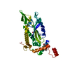 8a1bC  3l6tS C: citing same article ( S: Starting model for refinement |
|---|---|
| Similar structure data |
-Links
 PDBj
PDBj


- Assembly
| Deposited unit | 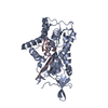
| ||||||||||
|---|---|---|---|---|---|---|---|---|---|---|---|
| 1 |
| ||||||||||
| Unit cell |
|
-Components
| #1: Protein | Mass: 34752.801 Da / Num. of mol.: 1 Source method: isolated from a genetically manipulated source Source: (gene. exp.) |
|---|---|
| #2: DNA chain | Mass: 3420.231 Da / Num. of mol.: 1 / Source method: obtained synthetically / Source: (synth.) synthetic construct (others) |
| #3: Chemical | ChemComp-MN /   Mass: 54.938 Da / Num. of mol.: 1 / Source method: obtained synthetically / Formula: Mn / Feature type: SUBJECT OF INVESTIGATION Mass: 54.938 Da / Num. of mol.: 1 / Source method: obtained synthetically / Formula: Mn / Feature type: SUBJECT OF INVESTIGATION |
| #4: Water | ChemComp-HOH /  Mass: 18.015 Da / Num. of mol.: 36 / Source method: isolated from a natural source / Formula: H2O Mass: 18.015 Da / Num. of mol.: 36 / Source method: isolated from a natural source / Formula: H2O |
| Has ligand of interest | Y |
-Experimental details
-Experiment
| Experiment | Method: X-RAY DIFFRACTION / Number of used crystals: 1 |
|---|
- Sample preparation
| Crystal | Density Matthews: 2.11 Å3/Da / Density % sol: 41.67 % |
|---|---|
| Crystal grow | Temperature: 293 K / Method: vapor diffusion, sitting drop Details: 10% PEG 20000, 20% PEG MME 550, 0.3 M sodium nitrate, 0.03 M disodium hydrogen phosphate, 0.03 M ammonium sulfate, 0.1 M MES/imidazole pH 6.5 |
-Data collection
| Diffraction | Mean temperature: 100 K / Serial crystal experiment: N |
|---|---|
| Diffraction source | Source: SYNCHROTRON / Site: ESRF  / Beamline: ID30B / Wavelength: 0.974 Å / Beamline: ID30B / Wavelength: 0.974 Å |
| Detector | Type: DECTRIS PILATUS 6M / Detector: PIXEL / Date: Feb 27, 2021 |
| Radiation | Protocol: SINGLE WAVELENGTH / Monochromatic (M) / Laue (L): M / Scattering type: x-ray |
| Radiation wavelength | Wavelength: 0.974 Å / Relative weight: 1 |
| Reflection | Resolution: 2.1→40.82 Å / Num. obs: 19496 / % possible obs: 99.85 % / Redundancy: 2 % / Biso Wilson estimate: 41.44 Å2 / CC1/2: 0.996 / Net I/σ(I): 9.16 |
| Reflection shell | Resolution: 2.1→2.175 Å / Redundancy: 2 % / Num. unique obs: 1908 / CC1/2: 0.751 / % possible all: 99.79 |
- Processing
| Software |
| ||||||||||||||||||||||||||||||||||||||||||||||||
|---|---|---|---|---|---|---|---|---|---|---|---|---|---|---|---|---|---|---|---|---|---|---|---|---|---|---|---|---|---|---|---|---|---|---|---|---|---|---|---|---|---|---|---|---|---|---|---|---|---|
| Refinement | Method to determine structure: MOLECULAR REPLACEMENT Starting model: 3L6T Resolution: 2.1→40.82 Å / SU ML: 0.26 / Cross valid method: THROUGHOUT / σ(F): 1.35 / Phase error: 31.83 / Stereochemistry target values: ML
| ||||||||||||||||||||||||||||||||||||||||||||||||
| Solvent computation | Shrinkage radii: 0.9 Å / VDW probe radii: 1.11 Å / Solvent model: FLAT BULK SOLVENT MODEL | ||||||||||||||||||||||||||||||||||||||||||||||||
| Displacement parameters | Biso max: 214.49 Å2 / Biso mean: 67.1356 Å2 / Biso min: 30.48 Å2 | ||||||||||||||||||||||||||||||||||||||||||||||||
| Refinement step | Cycle: final / Resolution: 2.1→40.82 Å
| ||||||||||||||||||||||||||||||||||||||||||||||||
| LS refinement shell | Refine-ID: X-RAY DIFFRACTION / Rfactor Rfree error: 0 / Total num. of bins used: 7 / % reflection obs: 100 %
| ||||||||||||||||||||||||||||||||||||||||||||||||
| Refinement TLS params. | Method: refined / Origin x: 3.3439 Å / Origin y: 10.8771 Å / Origin z: 22.3112 Å
| ||||||||||||||||||||||||||||||||||||||||||||||||
| Refinement TLS group |
|
