 Movie
Movie Controller

[English] 日本語
 Yorodumi
- PDB-8a19: Structure of a leucinostatin derivative determined by host lattic... -
+ Open data

- Basic information
| Entry | Database: PDB / ID: 8a19 | ||||||
|---|---|---|---|---|---|---|---|
| Title | Structure of a leucinostatin derivative determined by host lattice display : L1E4V1 construct | ||||||
 Components Components |
| ||||||
 Keywords Keywords | DE NOVO PROTEIN / Host lattice display / leucinostatin / EngBF / DARPin | ||||||
| Function / homology | Beta-galactosidase; Chain A, domain 5 - #10 / Beta-galactosidase; Chain A, domain 5 / Distorted Sandwich / Mainly Beta / :  Function and homology information Function and homology information | ||||||
| Biological species | synthetic construct (others) | ||||||
| Method |  X-RAY DIFFRACTION / SYNCHROTRON / FOURIER SYNTHESIS / Resolution: 2.358 Å X-RAY DIFFRACTION / SYNCHROTRON / FOURIER SYNTHESIS / Resolution: 2.358 Å | ||||||
 Authors Authors | Mittl, P.R.E. | ||||||
| Funding support | 1items
| ||||||
 Citation Citation | Journal: Acta Crystallogr D Struct Biol / Year: 2022 Title: Structure of a hydrophobic leucinostatin derivative determined by host lattice display. Authors: Kiss, C. / Gall, F.M. / Dreier, B. / Adams, M. / Riedl, R. / Pluckthun, A. / Mittl, P.R.E. | ||||||
| History |
|
- Structure visualization
| Structure viewer | Molecule: MolmilJmol/JSmol |
|---|
- Downloads & links
-Download
| PDBx/mmCIF format | 8a19.cif.gz | 314 KB | Display | PDBx/mmCIF format |
|---|---|---|---|---|
| PDB format | pdb8a19.ent.gz | 240.9 KB | Display | PDB format |
| PDBx/mmJSON format | 8a19.json.gz | Tree view | PDBx/mmJSON format | |
| Others |  Other downloads Other downloads |
-Validation report
| Arichive directory | https://data.pdbj.org/pub/pdb/validation_reports/a1/8a19ftp://data.pdbj.org/pub/pdb/validation_reports/a1/8a19 | HTTPS FTP |
|---|
-Related structure data
| Related structure data | 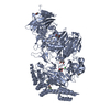 8a1aC 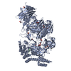 6qfkS S: Starting model for refinement C: citing same article ( |
|---|---|
| Similar structure data |
-Links
 PDBj
PDBj
- Assembly
| Deposited unit | 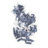
| ||||||||
|---|---|---|---|---|---|---|---|---|---|
| 1 |
| ||||||||
| Unit cell |
|
-Components
-Protein / Protein/peptide , 2 types, 2 molecules AB
| #1: Protein | Mass: 146990.641 Da / Num. of mol.: 1 Source method: isolated from a genetically manipulated source Details: Host lattice / Source: (gene. exp.) synthetic construct (others) / Production host:  |
|---|---|
| #2: Protein/peptide | Mass: 1174.493 Da / Num. of mol.: 1 / Source method: obtained synthetically / Source: (synth.) synthetic construct (others) |
-Non-polymers , 5 types, 1265 molecules 


| #3: Chemical | ChemComp-MN /  Mass: 54.938 Da / Num. of mol.: 4 / Source method: obtained synthetically / Formula: Mn Mass: 54.938 Da / Num. of mol.: 4 / Source method: obtained synthetically / Formula: Mn#4: Chemical | ChemComp-MPD / (  Mass: 118.174 Da / Num. of mol.: 4 / Source method: obtained synthetically / Formula: C6H14O2 / Comment: precipitant*YM Mass: 118.174 Da / Num. of mol.: 4 / Source method: obtained synthetically / Formula: C6H14O2 / Comment: precipitant*YM#5: Chemical | ChemComp-MES / |  Mass: 195.237 Da / Num. of mol.: 1 / Source method: obtained synthetically / Formula: C6H13NO4S / Comment: pH buffer*YM Mass: 195.237 Da / Num. of mol.: 1 / Source method: obtained synthetically / Formula: C6H13NO4S / Comment: pH buffer*YM#6: Chemical | ChemComp-CL /  Mass: 35.453 Da / Num. of mol.: 5 / Source method: obtained synthetically / Formula: Cl Mass: 35.453 Da / Num. of mol.: 5 / Source method: obtained synthetically / Formula: Cl#7: Water | ChemComp-HOH / | Mass: 18.015 Da / Num. of mol.: 1251 / Source method: isolated from a natural source / Formula: H2O |
|---|
-Details
| Has ligand of interest | N |
|---|
-Experimental details
-Experiment
| Experiment | Method: X-RAY DIFFRACTION / Number of used crystals: 1 |
|---|
- Sample preparation
| Crystal | Density Matthews: 4.44 Å3/Da / Density % sol: 72.31 % |
|---|---|
| Crystal grow | Temperature: 277 K / Method: vapor diffusion / pH: 6.43 Details: 2.55 % PEG20000, 26.72 % MPD, 0.2M NaCl, 0.01M MnCl2, 0.1M MES, pH 6.43 |
-Data collection
| Diffraction | Mean temperature: 100 K / Serial crystal experiment: N |
|---|---|
| Diffraction source | Source: SYNCHROTRON / Site: SLS  / Beamline: X06SA / Wavelength: 1 Å / Beamline: X06SA / Wavelength: 1 Å |
| Detector | Type: DECTRIS EIGER X 16M / Detector: PIXEL / Date: Mar 7, 2021 |
| Radiation | Protocol: SINGLE WAVELENGTH / Monochromatic (M) / Laue (L): M / Scattering type: x-ray |
| Radiation wavelength | Wavelength: 1 Å / Relative weight: 1 |
| Reflection | Resolution: 2.36→98 Å / Num. obs: 96740 / % possible obs: 94.8 % / Redundancy: 11.9 % / CC1/2: 0.996 / Rmerge(I) obs: 0.227 / Rpim(I) all: 0.069 / Rrim(I) all: 0.237 / Net I/σ(I): 8.8 |
| Reflection shell | Resolution: 2.36→2.47 Å / Num. unique obs: 4841 / CC1/2: 0.599 / Rpim(I) all: 0.575 / % possible all: 51 |
- Processing
| Software |
| ||||||||||||||||||||||||||||||||||||||||||||||||||||||||||||
|---|---|---|---|---|---|---|---|---|---|---|---|---|---|---|---|---|---|---|---|---|---|---|---|---|---|---|---|---|---|---|---|---|---|---|---|---|---|---|---|---|---|---|---|---|---|---|---|---|---|---|---|---|---|---|---|---|---|---|---|---|---|
| Refinement | Method to determine structure: FOURIER SYNTHESIS Starting model: 6QFK Resolution: 2.358→34.52 Å / Cor.coef. Fo:Fc: 0.96 / Cor.coef. Fo:Fc free: 0.948 / SU R Cruickshank DPI: 0.194 / Cross valid method: THROUGHOUT / SU R Blow DPI: 0.206 / SU Rfree Blow DPI: 0.167 / SU Rfree Cruickshank DPI: 0.164
| ||||||||||||||||||||||||||||||||||||||||||||||||||||||||||||
| Displacement parameters | Biso mean: 51.85 Å2
| ||||||||||||||||||||||||||||||||||||||||||||||||||||||||||||
| Refine analyze | Luzzati coordinate error obs: 0.24 Å | ||||||||||||||||||||||||||||||||||||||||||||||||||||||||||||
| Refinement step | Cycle: LAST / Resolution: 2.358→34.52 Å
| ||||||||||||||||||||||||||||||||||||||||||||||||||||||||||||
| Refine LS restraints |
| ||||||||||||||||||||||||||||||||||||||||||||||||||||||||||||
| LS refinement shell | Resolution: 2.36→2.42 Å
|
