 Movie
Movie Controller

[English] 日本語
 Yorodumi
- PDB-8a0d: Crystal structure of the major guinea pig allergen Cav p 1.0101 p... -
+ Open data

- Basic information
| Entry | Database: PDB / ID: 8a0d | ||||||
|---|---|---|---|---|---|---|---|
| Title | Crystal structure of the major guinea pig allergen Cav p 1.0101 part of the lipocalin family | ||||||
 Components Components | Allergen lipocalin Cav p 1 isoform 1 | ||||||
 Keywords Keywords | ALLERGEN / mammalian respiratory allergens | ||||||
| Function / homology | Lipocalin, OBP-like / odorant binding / Lipocalin / small molecule binding / Lipocalin / cytosolic fatty-acid binding protein family / Lipocalin/cytosolic fatty-acid binding domain / Calycin / extracellular space / Allergen lipocalin Cav p 1 isoform 1 Function and homology information Function and homology information | ||||||
| Biological species |  Cavia porcellus (domestic guinea pig) Cavia porcellus (domestic guinea pig) | ||||||
| Method |  X-RAY DIFFRACTION / SYNCHROTRON / MOLECULAR REPLACEMENT / Resolution: 3.685 Å X-RAY DIFFRACTION / SYNCHROTRON / MOLECULAR REPLACEMENT / Resolution: 3.685 Å | ||||||
 Authors Authors | Herman, R. / Charlier, P. / Janssen-Weets, B. / Hilger, C. / Swiontek, K. | ||||||
| Funding support |  Belgium, 1items Belgium, 1items
| ||||||
 Citation Citation | Journal: Front Allergy / Year: 2022 Title: Mammalian derived lipocalin and secretoglobin respiratory allergens strongly bind ligands with potentially immune modulating properties. Authors: Janssen-Weets, B. / Kerff, F. / Swiontek, K. / Kler, S. / Czolk, R. / Revets, D. / Kuehn, A. / Bindslev-Jensen, C. / Ollert, M. / Hilger, C. | ||||||
| History |
|
- Structure visualization
| Structure viewer | Molecule: MolmilJmol/JSmol |
|---|
- Downloads & links
-Download
| PDBx/mmCIF format | 8a0d.cif.gz | 266.5 KB | Display | PDBx/mmCIF format |
|---|---|---|---|---|
| PDB format | pdb8a0d.ent.gz | 224.4 KB | Display | PDB format |
| PDBx/mmJSON format | 8a0d.json.gz | Tree view | PDBx/mmJSON format | |
| Others |  Other downloads Other downloads |
-Validation report
| Summary document | 8a0d_validation.pdf.gz | 451.8 KB | Display | wwPDB validaton report |
|---|---|---|---|---|
| Full document | 8a0d_full_validation.pdf.gz | 454.3 KB | Display | |
| Data in XML | 8a0d_validation.xml.gz | 25.5 KB | Display | |
| Data in CIF | 8a0d_validation.cif.gz | 31.6 KB | Display | |
| Arichive directory | https://data.pdbj.org/pub/pdb/validation_reports/a0/8a0dftp://data.pdbj.org/pub/pdb/validation_reports/a0/8a0d | HTTPS FTP |
-Related structure data
| Related structure data | 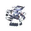 1e5pS S: Starting model for refinement |
|---|---|
| Similar structure data |
-Links
 PDBj
PDBj

- Assembly
| Deposited unit | 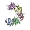
| ||||||||
|---|---|---|---|---|---|---|---|---|---|
| 1 | 
| ||||||||
| 2 | 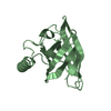
| ||||||||
| 3 | 
| ||||||||
| 4 | 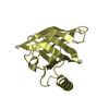
| ||||||||
| 5 | 
| ||||||||
| Unit cell |
|
-Components
| #1: Protein | Mass: 17806.502 Da / Num. of mol.: 5 Source method: isolated from a genetically manipulated source Source: (gene. exp.) Cavia porcellus (domestic guinea pig) / Gene: lcn / Production host:  Has protein modification | Y | |
|---|
-Experimental details
-Experiment
| Experiment | Method: X-RAY DIFFRACTION / Number of used crystals: 1 |
|---|
- Sample preparation
| Crystal | Density Matthews: 3.17 Å3/Da / Density % sol: 61.17 % |
|---|---|
| Crystal grow | Temperature: 293 K / Method: vapor diffusion, sitting drop / pH: 5 Details: 250 nl protein 4 mg/mL Tris-HCl 20 mM pH 7, 250 nl polyvinylpyrrolidone K15 50% w/v, citrate 0.1M pH 5, 50 nL of 6-Aminohexanoic acid 30% w/v and 50 nL of 1-decanol 1 mM in ethanol 1% v/v |
-Data collection
| Diffraction | Mean temperature: 100 K / Serial crystal experiment: N |
|---|---|
| Diffraction source | Source: SYNCHROTRON / Site: SOLEIL  / Beamline: PROXIMA 1 / Wavelength: 0.97857 Å / Beamline: PROXIMA 1 / Wavelength: 0.97857 Å |
| Detector | Type: DECTRIS PILATUS 6M / Detector: PIXEL / Date: Nov 1, 2014 |
| Radiation | Protocol: SINGLE WAVELENGTH / Monochromatic (M) / Laue (L): M / Scattering type: x-ray |
| Radiation wavelength | Wavelength: 0.97857 Å / Relative weight: 1 |
| Reflection | Resolution: 3.685→47.5 Å / Num. obs: 12775 / % possible obs: 99.9 % / Redundancy: 13.3 % / CC1/2: 0.992 / Rmerge(I) obs: 0.272 / Net I/σ(I): 10.2 |
| Reflection shell | Resolution: 3.7→3.78 Å / Redundancy: 12 % / Rmerge(I) obs: 0.877 / Mean I/σ(I) obs: 2.7 / Num. unique obs: 922 / CC1/2: 0.87 / % possible all: 100 |
- Processing
| Software |
| ||||||||||||||||||||||||||||||||||||||||||||||||||||||||||||
|---|---|---|---|---|---|---|---|---|---|---|---|---|---|---|---|---|---|---|---|---|---|---|---|---|---|---|---|---|---|---|---|---|---|---|---|---|---|---|---|---|---|---|---|---|---|---|---|---|---|---|---|---|---|---|---|---|---|---|---|---|---|
| Refinement | Method to determine structure: MOLECULAR REPLACEMENT Starting model: 1E5P Resolution: 3.685→29.91 Å / Cor.coef. Fo:Fc: 0.869 / Cor.coef. Fo:Fc free: 0.871 / Cross valid method: THROUGHOUT / SU Rfree Blow DPI: 0.642
| ||||||||||||||||||||||||||||||||||||||||||||||||||||||||||||
| Displacement parameters | Biso mean: 94.47 Å2
| ||||||||||||||||||||||||||||||||||||||||||||||||||||||||||||
| Refine analyze | Luzzati coordinate error obs: 0.55 Å | ||||||||||||||||||||||||||||||||||||||||||||||||||||||||||||
| Refinement step | Cycle: LAST / Resolution: 3.685→29.91 Å
| ||||||||||||||||||||||||||||||||||||||||||||||||||||||||||||
| Refine LS restraints |
| ||||||||||||||||||||||||||||||||||||||||||||||||||||||||||||
| LS refinement shell | Resolution: 3.69→3.73 Å
|
